 Movie
Movie Controller


+ Open data

- Basic information
| Entry | Database: PDB / ID: 7en6 | ||||||
|---|---|---|---|---|---|---|---|
| Title | The crystal structure of Escherichia coli MurR in apo form | ||||||
 Components Components | HTH-type transcriptional regulator MurR | ||||||
 Keywords Keywords | GENE REGULATION / regulator / Apo-form / sugar-binding | ||||||
| Function / homology | Glucose-6-phosphate isomerase like protein; domain 1 / Rossmann fold / 3-Layer(aba) Sandwich / Alpha Beta / PHOSPHATE ION / :  Function and homology information Function and homology information | ||||||
| Biological species |  | ||||||
| Method |  X-RAY DIFFRACTION / SYNCHROTRON / MOLECULAR REPLACEMENT / molecular replacement / Resolution: 2.276 Å X-RAY DIFFRACTION / SYNCHROTRON / MOLECULAR REPLACEMENT / molecular replacement / Resolution: 2.276 Å | ||||||
 Authors Authors | Zhang, Y. / Chen, W. / Ji, Q. | ||||||
| Funding support |  China, 1items China, 1items
| ||||||
 Citation Citation | Journal: Nucleic Acids Res. / Year: 2022 Title: Molecular basis for cell-wall recycling regulation by transcriptional repressor MurR in Escherichia coli. Authors: Zhang, Y. / Chen, W. / Wu, D. / Liu, Y. / Wu, Z. / Li, J. / Zhang, S.Y. / Ji, Q. | ||||||
| History |
|
- Structure visualization
| Structure viewer | Molecule: MolmilJmol/JSmol |
|---|
- Downloads & links
-Download
| PDBx/mmCIF format | 7en6.cif.gz | 277 KB | Display | PDBx/mmCIF format |
|---|---|---|---|---|
| PDB format | pdb7en6.ent.gz | 224.7 KB | Display | PDB format |
| PDBx/mmJSON format | 7en6.json.gz | Tree view | PDBx/mmJSON format | |
| Others |  Other downloads Other downloads |
-Validation report
| Arichive directory | https://data.pdbj.org/pub/pdb/validation_reports/en/7en6ftp://data.pdbj.org/pub/pdb/validation_reports/en/7en6 | HTTPS FTP |
|---|
-Related structure data
| Related structure data | 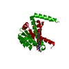 7en5C 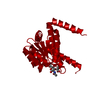 7en7C  4ivnS S: Starting model for refinement C: citing same article ( |
|---|---|
| Similar structure data |
-Links
 PDBj
PDBj- Assembly
| Deposited unit | 
| ||||||||
|---|---|---|---|---|---|---|---|---|---|
| 1 |
| ||||||||
| Unit cell |
|
-Components
| #1: Protein | Mass: 19652.518 Da / Num. of mol.: 4 Source method: isolated from a genetically manipulated source Source: (gene. exp.) #2: Chemical |   Mass: 94.971 Da / Num. of mol.: 2 / Source method: obtained synthetically / Formula: PO4 Mass: 94.971 Da / Num. of mol.: 2 / Source method: obtained synthetically / Formula: PO4#3: Water | ChemComp-HOH / |  Mass: 18.015 Da / Num. of mol.: 134 / Source method: isolated from a natural source / Formula: H2O Mass: 18.015 Da / Num. of mol.: 134 / Source method: isolated from a natural source / Formula: H2OHas ligand of interest | N | |
|---|
-Experimental details
-Experiment
| Experiment | Method: X-RAY DIFFRACTION / Number of used crystals: 1 |
|---|
- Sample preparation
| Crystal | Density Matthews: 2.23 Å3/Da / Density % sol: 44.86 % |
|---|---|
| Crystal grow | Temperature: 289 K / Method: vapor diffusion, sitting drop Details: 0.2 M L-proline, 0.1 M HEPES pH 7.5, 10% w/v polyethylene glycol 3,350 |
-Data collection
| Diffraction | Mean temperature: 100 K / Serial crystal experiment: N |
|---|---|
| Diffraction source | Source: SYNCHROTRON / Site: SSRF / Beamline: BL19U1 / Wavelength: 0.9785 Å |
| Detector | Type: AGILENT ATLAS CCD / Detector: CCD / Date: Oct 24, 2016 |
| Radiation | Protocol: SINGLE WAVELENGTH / Monochromatic (M) / Laue (L): M / Scattering type: x-ray |
| Radiation wavelength | Wavelength: 0.9785 Å / Relative weight: 1 |
| Reflection | Resolution: 2.27→50 Å / Num. obs: 32333 / % possible obs: 99.8 % / Redundancy: 12.5 % / Rmerge(I) obs: 0.079 / Rpim(I) all: 0.023 / Rrim(I) all: 0.082 / Net I/σ(I): 27.7 |
| Reflection shell | Resolution: 2.27→2.31 Å / Redundancy: 11.8 % / Rmerge(I) obs: 0.568 / Mean I/σ(I) obs: 2.6 / Num. unique obs: 1549 / CC1/2: 0.943 / CC star: 0.985 / Rpim(I) all: 0.169 / Rrim(I) all: 0.594 / % possible all: 99.8 |
-Phasing
| Phasing | Method: molecular replacement |
|---|
- Processing
| Software |
| |||||||||||||||||||||||||||||||||||||||||||||||||||||||||||||||||||||||||||||||||||||||||||||||||||||||||||||||||||||||||||||
|---|---|---|---|---|---|---|---|---|---|---|---|---|---|---|---|---|---|---|---|---|---|---|---|---|---|---|---|---|---|---|---|---|---|---|---|---|---|---|---|---|---|---|---|---|---|---|---|---|---|---|---|---|---|---|---|---|---|---|---|---|---|---|---|---|---|---|---|---|---|---|---|---|---|---|---|---|---|---|---|---|---|---|---|---|---|---|---|---|---|---|---|---|---|---|---|---|---|---|---|---|---|---|---|---|---|---|---|---|---|---|---|---|---|---|---|---|---|---|---|---|---|---|---|---|---|---|
| Refinement | Method to determine structure: MOLECULAR REPLACEMENT Starting model: 4IVN Resolution: 2.276→38.792 Å / SU ML: 0.29 / Cross valid method: THROUGHOUT / σ(F): 1.34 / Phase error: 29.89 / Stereochemistry target values: ML
| |||||||||||||||||||||||||||||||||||||||||||||||||||||||||||||||||||||||||||||||||||||||||||||||||||||||||||||||||||||||||||||
| Solvent computation | Shrinkage radii: 0.9 Å / VDW probe radii: 1.11 Å / Solvent model: FLAT BULK SOLVENT MODEL | |||||||||||||||||||||||||||||||||||||||||||||||||||||||||||||||||||||||||||||||||||||||||||||||||||||||||||||||||||||||||||||
| Displacement parameters | Biso max: 135.41 Å2 / Biso mean: 59.2619 Å2 / Biso min: 25.81 Å2 | |||||||||||||||||||||||||||||||||||||||||||||||||||||||||||||||||||||||||||||||||||||||||||||||||||||||||||||||||||||||||||||
| Refinement step | Cycle: final / Resolution: 2.276→38.792 Å
| |||||||||||||||||||||||||||||||||||||||||||||||||||||||||||||||||||||||||||||||||||||||||||||||||||||||||||||||||||||||||||||
| LS refinement shell | Refine-ID: X-RAY DIFFRACTION / Rfactor Rfree error: 0
| |||||||||||||||||||||||||||||||||||||||||||||||||||||||||||||||||||||||||||||||||||||||||||||||||||||||||||||||||||||||||||||
| Refinement TLS params. | Method: refined / Refine-ID: X-RAY DIFFRACTION
| |||||||||||||||||||||||||||||||||||||||||||||||||||||||||||||||||||||||||||||||||||||||||||||||||||||||||||||||||||||||||||||
| Refinement TLS group |
|
