 Movie
Movie Controller


+ Open data

- Basic information
| Entry | Database: PDB / ID: 7elf | ||||||
|---|---|---|---|---|---|---|---|
| Title | Nitrilase-Like Protein Nit2 from Kluyve-romyces lactis | ||||||
 Components Components | KLLA0E15247p | ||||||
 Keywords Keywords | HYDROLASE / Nitrilase / Nit2 | ||||||
| Function / homology | Uncharacterised protein family UPF0012, conserved site / Nit1/2, carbon-nitrogen hydrolase domain / Uncharacterized protein family UPF0012 signature. / Carbon-nitrogen hydrolase domain profile. / Carbon-nitrogen hydrolase superfamily / Carbon-nitrogen hydrolase / Carbon-nitrogen hydrolase / hydrolase activity, acting on carbon-nitrogen (but not peptide) bonds, in linear amides / KLLA0E15247p Function and homology information Function and homology information | ||||||
| Biological species |  Kluyveromyces lactis (yeast) Kluyveromyces lactis (yeast) | ||||||
| Method |  X-RAY DIFFRACTION / SYNCHROTRON / MOLECULAR REPLACEMENT / Resolution: 2.2 Å X-RAY DIFFRACTION / SYNCHROTRON / MOLECULAR REPLACEMENT / Resolution: 2.2 Å | ||||||
 Authors Authors | Jin, C.W. / Chang, J.H. | ||||||
| Funding support |  Korea, Republic Of, 1items Korea, Republic Of, 1items
| ||||||
 Citation Citation | Journal: Crystals / Year: 2021 Title: Crystal Structure of Nitrilase-Like Protein Nit2 from Kluyveromyces lactis. Authors: Jin, C.W. / Jin, H.S. / Jeong, B.C. / Cho, D.H. / Chun, H.S. / Kim, W.K. / Chang, J.H. | ||||||
| History |
|
- Structure visualization
| Structure viewer | Molecule: MolmilJmol/JSmol |
|---|
- Downloads & links
-Download
| PDBx/mmCIF format | 7elf.cif.gz | 458.4 KB | Display | PDBx/mmCIF format |
|---|---|---|---|---|
| PDB format | pdb7elf.ent.gz | 377.2 KB | Display | PDB format |
| PDBx/mmJSON format | 7elf.json.gz | Tree view | PDBx/mmJSON format | |
| Others |  Other downloads Other downloads |
-Validation report
| Arichive directory | https://data.pdbj.org/pub/pdb/validation_reports/el/7elfftp://data.pdbj.org/pub/pdb/validation_reports/el/7elf | HTTPS FTP |
|---|
-Related structure data
| Related structure data | 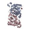 4h5uS S: Starting model for refinement |
|---|---|
| Similar structure data |
-Links
 PDBj
PDBj- Assembly
| Deposited unit | 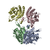
| |||||||||
|---|---|---|---|---|---|---|---|---|---|---|
| 1 | 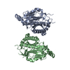
| |||||||||
| 2 | 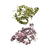
| |||||||||
| Unit cell |
| |||||||||
| Components on special symmetry positions |
|
-Components
| #1: Protein | Mass: 33633.520 Da / Num. of mol.: 4 Source method: isolated from a genetically manipulated source Source: (gene. exp.) Kluyveromyces lactis (strain ATCC 8585 / CBS 2359 / DSM 70799 / NBRC 1267 / NRRL Y-1140 / WM37) (yeast)Strain: ATCC 8585 / CBS 2359 / DSM 70799 / NBRC 1267 / NRRL Y-1140 / WM37 Gene: KLLA0_E15247g / Production host:  #2: Water | ChemComp-HOH / |  Mass: 18.015 Da / Num. of mol.: 543 / Source method: isolated from a natural source / Formula: H2O Mass: 18.015 Da / Num. of mol.: 543 / Source method: isolated from a natural source / Formula: H2O |
|---|
-Experimental details
-Experiment
| Experiment | Method: X-RAY DIFFRACTION / Number of used crystals: 1 |
|---|
- Sample preparation
| Crystal | Density Matthews: 2.57 Å3/Da / Density % sol: 52.06 % |
|---|---|
| Crystal grow | Temperature: 277 K / Method: vapor diffusion, hanging drop Details: 26% w/v polyethylene glycol (PEG 3350), 0.3M Ammonium tartrate dibasic |
-Data collection
| Diffraction | Mean temperature: 100 K / Serial crystal experiment: N |
|---|---|
| Diffraction source | Source: SYNCHROTRON / Site: PAL/PLS / Beamline: 5C (4A) / Wavelength: 1 Å |
| Detector | Type: ADSC QUANTUM 315 / Detector: CCD / Date: Mar 23, 2019 |
| Radiation | Protocol: SINGLE WAVELENGTH / Monochromatic (M) / Laue (L): M / Scattering type: x-ray |
| Radiation wavelength | Wavelength: 1 Å / Relative weight: 1 |
| Reflection | Resolution: 2.2→50 Å / Num. obs: 68559 / % possible obs: 99.9 % / Redundancy: 5.7 % / CC1/2: 0.991 / Net I/σ(I): 16.96 |
| Reflection shell | Resolution: 2.2→2.28 Å / Num. unique obs: 6799 / CC1/2: 0.862 |
- Processing
| Software |
| |||||||||||||||||||||||||||||||||||||||||||||||||||||||||||||||||||||||||||||||||||||||||||||||||||||||||
|---|---|---|---|---|---|---|---|---|---|---|---|---|---|---|---|---|---|---|---|---|---|---|---|---|---|---|---|---|---|---|---|---|---|---|---|---|---|---|---|---|---|---|---|---|---|---|---|---|---|---|---|---|---|---|---|---|---|---|---|---|---|---|---|---|---|---|---|---|---|---|---|---|---|---|---|---|---|---|---|---|---|---|---|---|---|---|---|---|---|---|---|---|---|---|---|---|---|---|---|---|---|---|---|---|---|---|
| Refinement | Method to determine structure: MOLECULAR REPLACEMENT Starting model: 4H5U Resolution: 2.2→45.68 Å / SU ML: 0.25 / Cross valid method: FREE R-VALUE / σ(F): 0 / Phase error: 23.01 / Stereochemistry target values: ML
| |||||||||||||||||||||||||||||||||||||||||||||||||||||||||||||||||||||||||||||||||||||||||||||||||||||||||
| Solvent computation | Shrinkage radii: 0.9 Å / VDW probe radii: 1.11 Å / Solvent model: FLAT BULK SOLVENT MODEL | |||||||||||||||||||||||||||||||||||||||||||||||||||||||||||||||||||||||||||||||||||||||||||||||||||||||||
| Refinement step | Cycle: LAST / Resolution: 2.2→45.68 Å
| |||||||||||||||||||||||||||||||||||||||||||||||||||||||||||||||||||||||||||||||||||||||||||||||||||||||||
| Refine LS restraints |
| |||||||||||||||||||||||||||||||||||||||||||||||||||||||||||||||||||||||||||||||||||||||||||||||||||||||||
| LS refinement shell |
| |||||||||||||||||||||||||||||||||||||||||||||||||||||||||||||||||||||||||||||||||||||||||||||||||||||||||
| Refinement TLS params. | Method: refined / Origin x: 0.7591 Å / Origin y: 5.2596 Å / Origin z: 20.527 Å
| |||||||||||||||||||||||||||||||||||||||||||||||||||||||||||||||||||||||||||||||||||||||||||||||||||||||||
| Refinement TLS group | Selection details: all |
