 Movie
Movie Controller

[English] 日本語
 Yorodumi
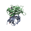
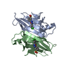
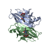
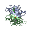
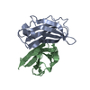
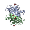
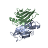
- PDB-7ejr: Crystal structure of V30M mutated transthyretin in complex with 8... -
+ Open data

- Basic information
| Entry | Database: PDB / ID: 7ejr | ||||||
|---|---|---|---|---|---|---|---|
| Title | Crystal structure of V30M mutated transthyretin in complex with 8-chloro-9-oxo-9H-xanthene-3-carboxylic acid | ||||||
 Components Components | Transthyretin | ||||||
 Keywords Keywords | TRANSPORT PROTEIN / amyloidosis / inhibitor / complex | ||||||
| Function / homology |  Function and homology information Function and homology informationDefective visual phototransduction due to STRA6 loss of function / negative regulation of glomerular filtration / The canonical retinoid cycle in rods (twilight vision) / purine nucleobase metabolic process / hormone binding / Non-integrin membrane-ECM interactions / phototransduction, visible light / molecular sequestering activity / retinoid metabolic process / Retinoid metabolism and transport ...Defective visual phototransduction due to STRA6 loss of function / negative regulation of glomerular filtration / The canonical retinoid cycle in rods (twilight vision) / purine nucleobase metabolic process / hormone binding / Non-integrin membrane-ECM interactions / phototransduction, visible light / molecular sequestering activity / retinoid metabolic process / Retinoid metabolism and transport / hormone activity / azurophil granule lumen / Amyloid fiber formation / Neutrophil degranulation / protein-containing complex binding / protein-containing complex / : / extracellular exosome / extracellular region / identical protein binding Similarity search - Function | ||||||
| Biological species |  Homo sapiens (human) Homo sapiens (human) | ||||||
| Method |  X-RAY DIFFRACTION / SYNCHROTRON / MOLECULAR REPLACEMENT / Resolution: 1.451 Å X-RAY DIFFRACTION / SYNCHROTRON / MOLECULAR REPLACEMENT / Resolution: 1.451 Å | ||||||
 Authors Authors | Kitakami, R. / Yokoyama, T. / Mizuguchi, M. | ||||||
 Citation Citation | Journal: Bioorg.Med.Chem. / Year: 2021 Title: Inhibitory activities of anthraquinone and xanthone derivatives against transthyretin amyloidogenesis. Authors: Kitakami, R. / Inui, K. / Nakagawa, Y. / Sawai, Y. / Katayama, W. / Yokoyama, T. / Okada, T. / Kanamitsu, K. / Nakagawa, S. / Toyooka, N. / Mizuguchi, M. | ||||||
| History |
|
- Structure visualization
| Structure viewer | Molecule: MolmilJmol/JSmol |
|---|
- Downloads & links
-Download
| PDBx/mmCIF format | 7ejr.cif.gz | 66.1 KB | Display | PDBx/mmCIF format |
|---|---|---|---|---|
| PDB format | pdb7ejr.ent.gz | 46.2 KB | Display | PDB format |
| PDBx/mmJSON format | 7ejr.json.gz | Tree view | PDBx/mmJSON format | |
| Others |  Other downloads Other downloads |
-Validation report
| Arichive directory | https://data.pdbj.org/pub/pdb/validation_reports/ej/7ejrftp://data.pdbj.org/pub/pdb/validation_reports/ej/7ejr | HTTPS FTP |
|---|
-Related structure data
| Related structure data |  7dt3C  7dt5C  7dt6C 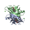 7dt8C 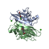 7ejqC 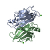 4n85S S: Starting model for refinement C: citing same article ( |
|---|---|
| Similar structure data |


-Links
 PDBj
PDBj


- Assembly
| Deposited unit | 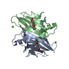
| ||||||||||||
|---|---|---|---|---|---|---|---|---|---|---|---|---|---|
| 1 |
| ||||||||||||
| Unit cell |
| ||||||||||||
| Components on special symmetry positions |
|
-Components
| #1: Protein | Mass: 17213.469 Da / Num. of mol.: 2 Source method: isolated from a genetically manipulated source Source: (gene. exp.) Homo sapiens (human) / Gene: TTR, PALB / Production host:  #2: Chemical |   Mass: 40.078 Da / Num. of mol.: 2 / Source method: obtained synthetically / Formula: Ca Mass: 40.078 Da / Num. of mol.: 2 / Source method: obtained synthetically / Formula: Ca#3: Chemical | 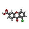  Mass: 274.656 Da / Num. of mol.: 2 / Source method: obtained synthetically / Formula: C14H7ClO4 / Feature type: SUBJECT OF INVESTIGATION Mass: 274.656 Da / Num. of mol.: 2 / Source method: obtained synthetically / Formula: C14H7ClO4 / Feature type: SUBJECT OF INVESTIGATION#4: Water | ChemComp-HOH / |  Mass: 18.015 Da / Num. of mol.: 175 / Source method: isolated from a natural source / Formula: H2O Mass: 18.015 Da / Num. of mol.: 175 / Source method: isolated from a natural source / Formula: H2OHas ligand of interest | Y | |
|---|
-Experimental details
-Experiment
| Experiment | Method: X-RAY DIFFRACTION / Number of used crystals: 1 |
|---|
- Sample preparation
| Crystal | Density Matthews: 1.65 Å3/Da / Density % sol: 25.58 % |
|---|---|
| Crystal grow | Temperature: 293 K / Method: vapor diffusion, hanging drop / Details: PEG400, CaCl2, NaOAc |
-Data collection
| Diffraction | Mean temperature: 100 K / Serial crystal experiment: N |
|---|---|
| Diffraction source | Source: SYNCHROTRON / Site: Photon Factory  / Beamline: BL-17A / Wavelength: 0.98 Å / Beamline: BL-17A / Wavelength: 0.98 Å |
| Detector | Type: DECTRIS PILATUS 6M / Detector: PIXEL / Date: Nov 29, 2018 |
| Radiation | Protocol: SINGLE WAVELENGTH / Monochromatic (M) / Laue (L): M / Scattering type: x-ray |
| Radiation wavelength | Wavelength: 0.98 Å / Relative weight: 1 |
| Reflection | Resolution: 1.45→35.47 Å / Num. obs: 41002 / % possible obs: 99.6 % / Redundancy: 6.5 % / CC1/2: 0.999 / Rpim(I) all: 0.018 / Rrim(I) all: 0.046 / Net I/σ(I): 21 |
| Reflection shell | Resolution: 1.45→1.5 Å / Mean I/σ(I) obs: 3.3 / Num. unique obs: 3921 / CC1/2: 0.915 / Rpim(I) all: 0.192 / Rrim(I) all: 0.497 |
- Processing
| Software |
| ||||||||||||||||||||||||||||||||||||||||||||||||||||||||||||||||||||||||||||||||||||||||||||||||||||||||||||||||
|---|---|---|---|---|---|---|---|---|---|---|---|---|---|---|---|---|---|---|---|---|---|---|---|---|---|---|---|---|---|---|---|---|---|---|---|---|---|---|---|---|---|---|---|---|---|---|---|---|---|---|---|---|---|---|---|---|---|---|---|---|---|---|---|---|---|---|---|---|---|---|---|---|---|---|---|---|---|---|---|---|---|---|---|---|---|---|---|---|---|---|---|---|---|---|---|---|---|---|---|---|---|---|---|---|---|---|---|---|---|---|---|---|---|
| Refinement | Method to determine structure: MOLECULAR REPLACEMENT Starting model: 4N85 Resolution: 1.451→31.429 Å / SU ML: 0.14 / Cross valid method: FREE R-VALUE / σ(F): 1.38 / Phase error: 20.34 / Stereochemistry target values: ML
| ||||||||||||||||||||||||||||||||||||||||||||||||||||||||||||||||||||||||||||||||||||||||||||||||||||||||||||||||
| Solvent computation | Shrinkage radii: 0.9 Å / VDW probe radii: 1.11 Å / Solvent model: FLAT BULK SOLVENT MODEL | ||||||||||||||||||||||||||||||||||||||||||||||||||||||||||||||||||||||||||||||||||||||||||||||||||||||||||||||||
| Refinement step | Cycle: LAST / Resolution: 1.451→31.429 Å
| ||||||||||||||||||||||||||||||||||||||||||||||||||||||||||||||||||||||||||||||||||||||||||||||||||||||||||||||||
| Refine LS restraints |
| ||||||||||||||||||||||||||||||||||||||||||||||||||||||||||||||||||||||||||||||||||||||||||||||||||||||||||||||||
| LS refinement shell |
|
