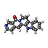
+ Open data
- Basic information
| Entry | Database: PDB / ID: 7eg4 | |||||||||||||||||||||||||||
|---|---|---|---|---|---|---|---|---|---|---|---|---|---|---|---|---|---|---|---|---|---|---|---|---|---|---|---|---|
| Title | Cryo-EM structure of nauclefine-induced PDE3A-SLFN12 complex | |||||||||||||||||||||||||||
 Components Components |
| |||||||||||||||||||||||||||
 Keywords Keywords | APOPTOSIS / nauclefine | |||||||||||||||||||||||||||
| Function / homology |  Function and homology information Function and homology information3',5'-cGMP-inhibited cyclic-nucleotide phosphodiesterase activity / estrogen binding / positive regulation of oocyte development / regulation of ribonuclease activity / regulation of meiotic nuclear division / cellular response to cGMP / rRNA catabolic process / negative regulation of adenylate cyclase-activating G protein-coupled receptor signaling pathway / positive regulation of vascular permeability / negative regulation of vascular permeability ...3',5'-cGMP-inhibited cyclic-nucleotide phosphodiesterase activity / estrogen binding / positive regulation of oocyte development / regulation of ribonuclease activity / regulation of meiotic nuclear division / cellular response to cGMP / rRNA catabolic process / negative regulation of adenylate cyclase-activating G protein-coupled receptor signaling pathway / positive regulation of vascular permeability / negative regulation of vascular permeability / negative regulation of cAMP/PKA signal transduction / oocyte maturation / : / 3',5'-cyclic-nucleotide phosphodiesterase / nuclear estrogen receptor activity / 3',5'-cyclic-nucleotide phosphodiesterase activity / 3',5'-cyclic-GMP phosphodiesterase activity / 3',5'-cyclic-AMP phosphodiesterase activity / RNA nuclease activity / cellular response to transforming growth factor beta stimulus / : / apoptotic signaling pathway / lipid metabolic process / ribosome binding / G alpha (s) signalling events / Hydrolases; Acting on ester bonds / G protein-coupled receptor signaling pathway / response to xenobiotic stimulus / negative regulation of apoptotic process / metal ion binding / nucleus / membrane / cytosol Similarity search - Function | |||||||||||||||||||||||||||
| Biological species |  Homo sapiens (human) Homo sapiens (human) | |||||||||||||||||||||||||||
| Method | ELECTRON MICROSCOPY / single particle reconstruction / cryo EM / Resolution: 3.2 Å | |||||||||||||||||||||||||||
 Authors Authors | Liu, N. / Chen, J. / Wang, X.D. / Wang, H.W. | |||||||||||||||||||||||||||
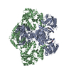
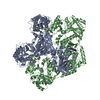
 Citation Citation |  Journal: Nat Commun / Year: 2021 Journal: Nat Commun / Year: 2021Title: Structure of PDE3A-SLFN12 complex and structure-based design for a potent apoptosis inducer of tumor cells. Authors: Jie Chen / Nan Liu / Yinpin Huang / Yuanxun Wang / Yuxing Sun / Qingcui Wu / Dianrong Li / Shuanhu Gao / Hong-Wei Wang / Niu Huang / Xiangbing Qi / Xiaodong Wang /  Abstract: Molecular glues are a class of small molecular drugs that mediate protein-protein interactions, that induce either the degradation or stabilization of target protein. A structurally diverse group of ...Molecular glues are a class of small molecular drugs that mediate protein-protein interactions, that induce either the degradation or stabilization of target protein. A structurally diverse group of chemicals, including 17-β-estradiol (E2), anagrelide, nauclefine, and DNMDP, induces apoptosis by forming complexes with phosphodiesterase 3A (PDE3A) and Schlafen 12 protein (SLFN12). They do so by binding to the PDE3A enzymatic pocket that allows the compound-bound PDE3A to recruit and stabilize SLFN12, which in turn blocks protein translation, leading to apoptosis. In this work, we report the high-resolution cryo-electron microscopy structure of PDE3A-SLFN12 complexes isolated from cultured HeLa cells pre-treated with either anagrelide, or nauclefine, or DNMDP. The PDE3A-SLFN12 complexes exhibit a butterfly-like shape, forming a heterotetramer with these small molecules, which are packed in a shallow pocket in the catalytic domain of PDE3A. The resulting small molecule-modified interface binds to the short helix (E552-I558) of SLFN12 through hydrophobic interactions, thus "gluing" the two proteins together. Based on the complex structure, we designed and synthesized analogs of anagrelide, a known drug used for the treatment of thrombocytosis, to enhance their interactions with SLFN12, and achieved superior efficacy in inducing apoptosis in cultured cells as well as in tumor xenografts. | |||||||||||||||||||||||||||
| History |
|
- Structure visualization
| Movie |
 Movie viewer Movie viewer |
|---|---|
| Structure viewer | Molecule:  Molmil Molmil Jmol/JSmol Jmol/JSmol |
- Downloads & links
- Download
| PDBx/mmCIF format |  7eg4.cif.gz 7eg4.cif.gz | 349.3 KB | Display |  PDBx/mmCIF format PDBx/mmCIF format |
|---|---|---|---|---|
| PDB format |  pdb7eg4.ent.gz pdb7eg4.ent.gz | 277.4 KB | Display |  PDB format PDB format |
| PDBx/mmJSON format |  7eg4.json.gz 7eg4.json.gz | Tree view |  PDBx/mmJSON format PDBx/mmJSON format | |
| Others |  Other downloads Other downloads |
-Validation report
| Arichive directory |  https://data.pdbj.org/pub/pdb/validation_reports/eg/7eg4 https://data.pdbj.org/pub/pdb/validation_reports/eg/7eg4 ftp://data.pdbj.org/pub/pdb/validation_reports/eg/7eg4 ftp://data.pdbj.org/pub/pdb/validation_reports/eg/7eg4 | HTTPS FTP |
|---|
-Related structure data
| Related structure data |  31105MC  7eg0C  7eg1C M: map data used to model this data C: citing same article ( |
|---|---|
| Similar structure data |
- Links
Links
- Assembly
| Deposited unit | 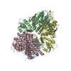
|
|---|---|
| 1 |
|
- Components
| #1: Protein | Mass: 49552.527 Da / Num. of mol.: 2 Source method: isolated from a genetically manipulated source Source: (gene. exp.)  Homo sapiens (human) / Gene: PDE3A / Cell line (production host): HEK293 / Production host: Homo sapiens (human) / Gene: PDE3A / Cell line (production host): HEK293 / Production host:  Homo sapiens (human) Homo sapiens (human)References: UniProt: Q14432, 3',5'-cyclic-nucleotide phosphodiesterase #2: Protein | Mass: 65665.656 Da / Num. of mol.: 2 / Mutation: K213R, S351A, D415S Source method: isolated from a genetically manipulated source Source: (gene. exp.)  Homo sapiens (human) / Gene: SLFN12 / Cell line (production host): HEK293 / Production host: Homo sapiens (human) / Gene: SLFN12 / Cell line (production host): HEK293 / Production host:  Homo sapiens (human) / References: UniProt: Q8IYM2 Homo sapiens (human) / References: UniProt: Q8IYM2#3: Chemical | #4: Chemical | ChemComp-MG / #5: Chemical | Has ligand of interest | N | Has protein modification | Y | |
|---|
-Experimental details
-Experiment
| Experiment | Method: ELECTRON MICROSCOPY |
|---|---|
| EM experiment | Aggregation state: PARTICLE / 3D reconstruction method: single particle reconstruction |
- Sample preparation
| Component |
| ||||||||||||||||||||||||
|---|---|---|---|---|---|---|---|---|---|---|---|---|---|---|---|---|---|---|---|---|---|---|---|---|---|
| Source (natural) |
| ||||||||||||||||||||||||
| Source (recombinant) |
| ||||||||||||||||||||||||
| Buffer solution | pH: 8 | ||||||||||||||||||||||||
| Specimen | Embedding applied: NO / Shadowing applied: NO / Staining applied: NO / Vitrification applied: YES | ||||||||||||||||||||||||
| Vitrification | Cryogen name: ETHANE |
- Electron microscopy imaging
| Experimental equipment |  Model: Titan Krios / Image courtesy: FEI Company |
|---|---|
| Microscopy | Model: FEI TITAN KRIOS |
| Electron gun | Electron source:  FIELD EMISSION GUN / Accelerating voltage: 300 kV / Illumination mode: FLOOD BEAM FIELD EMISSION GUN / Accelerating voltage: 300 kV / Illumination mode: FLOOD BEAM |
| Electron lens | Mode: BRIGHT FIELD |
| Image recording | Electron dose: 50 e/Å2 / Film or detector model: GATAN K3 (6k x 4k) |
- Processing
| Software | Name: PHENIX / Version: 1.14_3260: / Classification: refinement |
|---|---|
| EM software | Name: PHENIX / Category: model refinement |
| CTF correction | Type: PHASE FLIPPING ONLY |
| 3D reconstruction | Resolution: 3.2 Å / Resolution method: FSC 0.143 CUT-OFF / Num. of particles: 60683 / Symmetry type: POINT |
 Movie
Movie Controller


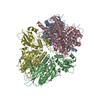
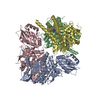




 PDBj
PDBj
