negative regulation of helicase activity / signal transduction by p53 class mediator / Loss of function of TP53 in cancer due to loss of tetramerization ability / Regulation of TP53 Expression / regulation of cell cycle G2/M phase transition / negative regulation of G1 to G0 transition / Transcriptional activation of cell cycle inhibitor p21 / negative regulation of pentose-phosphate shunt / Activation of NOXA and translocation to mitochondria / ATP-dependent DNA/DNA annealing activity ...negative regulation of helicase activity / signal transduction by p53 class mediator / Loss of function of TP53 in cancer due to loss of tetramerization ability / Regulation of TP53 Expression / regulation of cell cycle G2/M phase transition / negative regulation of G1 to G0 transition / Transcriptional activation of cell cycle inhibitor p21 / negative regulation of pentose-phosphate shunt / Activation of NOXA and translocation to mitochondria / ATP-dependent DNA/DNA annealing activity / oligodendrocyte apoptotic process / positive regulation of thymocyte apoptotic process / oxidative stress-induced premature senescence / bone marrow development / circadian behavior / cellular response to actinomycin D / positive regulation of programmed necrotic cell death / RUNX3 regulates CDKN1A transcription / TP53 Regulates Transcription of Death Receptors and Ligands / Activation of PUMA and translocation to mitochondria / TP53 regulates transcription of additional cell cycle genes whose exact role in the p53 pathway remain uncertain / mRNA transcription / Urea cycle / Regulation of TP53 Activity through Association with Co-factors / ER overload response / hematopoietic stem cell differentiation / Formation of Senescence-Associated Heterochromatin Foci (SAHF) / TP53 Regulates Transcription of Caspase Activators and Caspases / intrinsic apoptotic signaling pathway by p53 class mediator / entrainment of circadian clock by photoperiod / Zygotic genome activation (ZGA) / TP53 Regulates Transcription of Genes Involved in Cytochrome C Release / PI5P Regulates TP53 Acetylation / positive regulation of release of cytochrome c from mitochondria / hematopoietic progenitor cell differentiation / Association of TriC/CCT with target proteins during biosynthesis / negative regulation of telomere maintenance via telomerase / SUMOylation of transcription factors / TP53 regulates transcription of several additional cell death genes whose specific roles in p53-dependent apoptosis remain uncertain / intrinsic apoptotic signaling pathway in response to DNA damage by p53 class mediator / Transcriptional Regulation by VENTX / replicative senescence / TFIID-class transcription factor complex binding / intrinsic apoptotic signaling pathway in response to endoplasmic reticulum stress / viral process / positive regulation of intrinsic apoptotic signaling pathway / Pyroptosis / determination of adult lifespan / positive regulation of RNA polymerase II transcription preinitiation complex assembly / general transcription initiation factor binding / negative regulation of fibroblast proliferation / positive regulation of execution phase of apoptosis / type II interferon-mediated signaling pathway / TP53 Regulates Transcription of Genes Involved in G1 Cell Cycle Arrest / cellular response to glucose starvation / core promoter sequence-specific DNA binding / cis-regulatory region sequence-specific DNA binding / Regulation of TP53 Activity through Acetylation / intrinsic apoptotic signaling pathway / mitotic G1 DNA damage checkpoint signaling / response to gamma radiation / 14-3-3 protein binding / MDM2/MDM4 family protein binding / TP53 Regulates Transcription of Genes Involved in G2 Cell Cycle Arrest / protein phosphatase 2A binding / molecular function activator activity / transcription initiation-coupled chromatin remodeling / Regulation of PTEN gene transcription / tumor necrosis factor-mediated signaling pathway / cellular response to ionizing radiation / cellular response to xenobiotic stimulus / DNA damage response, signal transduction by p53 class mediator / TP53 Regulates Metabolic Genes / autophagy / TP53 Regulates Transcription of DNA Repair Genes / Regulation of NF-kappa B signaling / mRNA 3'-UTR binding / protein tetramerization / promoter-specific chromatin binding / Stabilization of p53 / molecular condensate scaffold activity / negative regulation of cell growth / cellular response to gamma radiation / nucleotide-excision repair / G2/M Checkpoints / receptor tyrosine kinase binding / Autodegradation of the E3 ubiquitin ligase COP1 / PML body / positive regulation of miRNA transcription / Oncogene Induced Senescence / DNA-binding transcription repressor activity, RNA polymerase II-specific / PKR-mediated signaling / Regulation of TP53 Activity through Methylation / G2/M DNA damage checkpoint / cellular senescence / DNA Damage/Telomere Stress Induced Senescence / Pre-NOTCH Transcription and Translation / positive regulation of reactive oxygen species metabolic process / intracellular protein localization / histone deacetylase binding Similarity search - Function
In the structure databanks used in Yorodumi, some data are registered as the other names, "COVID-19 virus" and "2019-nCoV". Here are the details of the virus and the list of structure data.
Jan 31, 2019. EMDB accession codes are about to change! (news from PDBe EMDB page)
EMDB accession codes are about to change! (news from PDBe EMDB page)
The allocation of 4 digits for EMDB accession codes will soon come to an end. Whilst these codes will remain in use, new EMDB accession codes will include an additional digit and will expand incrementally as the available range of codes is exhausted. The current 4-digit format prefixed with “EMD-” (i.e. EMD-XXXX) will advance to a 5-digit format (i.e. EMD-XXXXX), and so on. It is currently estimated that the 4-digit codes will be depleted around Spring 2019, at which point the 5-digit format will come into force.
The EM Navigator/Yorodumi systems omit the EMD- prefix.
Related info.:Q: What is EMD? / ID/Accession-code notation in Yorodumi/EM Navigator
Yorodumi is a browser for structure data from EMDB, PDB, SASBDB, etc.
This page is also the successor to EM Navigator detail page, and also detail information page/front-end page for Omokage search.
The word "yorodu" (or yorozu) is an old Japanese word meaning "ten thousand". "mi" (miru) is to see.
Related info.:EMDB / PDB / SASBDB / Comparison of 3 databanks / Yorodumi Search / Aug 31, 2016. New EM Navigator & Yorodumi / Yorodumi Papers / Jmol/JSmol / Function and homology information / Changes in new EM Navigator and Yorodumi
 Movie
Movie Controller

 Yorodumi
 Open data
 Basic information
 Components
 Keywords
 Function and homology information
 Homo sapiens (human)
 X-RAY DIFFRACTION /
  Authors
 China, 1items
  Citation
 Structure visualization
 Downloads & links
 Other downloads
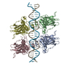
 PDBj
PDBj
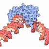

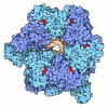

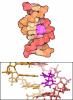

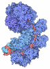
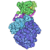


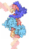
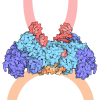
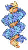
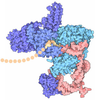
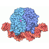
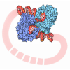
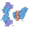
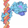
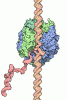
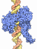
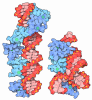



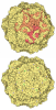
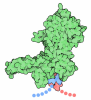
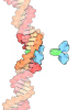
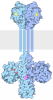

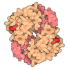
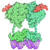
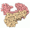

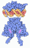

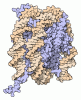
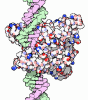
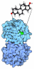

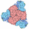
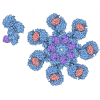

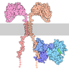

 Assembly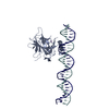

 Escherichia coli (E. coli) / References: UniProt: P04637


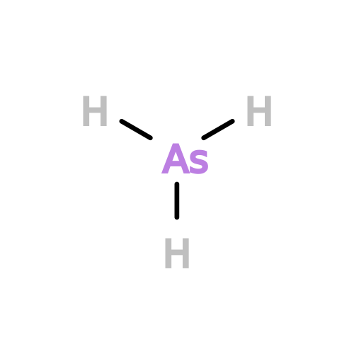 Mass: 74.922 Da / Num. of mol.: 1 / Source method: obtained synthetically / Formula: As / Feature type: SUBJECT OF INVESTIGATION
 Mass: 65.409 Da / Num. of mol.: 1 / Source method: obtained synthetically / Formula: Zn
 Sample preparation
 Processing