 Movie
Movie Controller

[English] 日本語
 Yorodumi
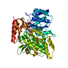
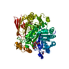
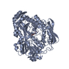
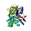
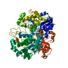
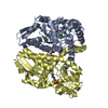
- PDB-7eaw: Trehalase of Arabidopsis thaliana acid mutant -D380A trehalose complex -
+ Open data

- Basic information
| Entry | Database: PDB / ID: 7eaw | ||||||
|---|---|---|---|---|---|---|---|
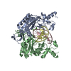
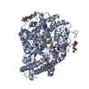
| Title | Trehalase of Arabidopsis thaliana acid mutant -D380A trehalose complex | ||||||
 Components Components | Trehalase | ||||||
 Keywords Keywords | HYDROLASE / trehalase / GH37 / Arabidopsis thaliana / Oryza sativa / trehalose / trehalose 6-phosphate / glycoside hydrolase / enzyme kinetics / enzyme structure | ||||||
| Function / homology |  Function and homology information Function and homology informationalpha,alpha-trehalase / alpha,alpha-trehalase activity / trehalose catabolic process / nucleus / plasma membrane / cytoplasm Similarity search - Function | ||||||
| Biological species |  | ||||||
| Method |  X-RAY DIFFRACTION / SYNCHROTRON / MOLECULAR REPLACEMENT / Resolution: 1.8 Å X-RAY DIFFRACTION / SYNCHROTRON / MOLECULAR REPLACEMENT / Resolution: 1.8 Å | ||||||
 Authors Authors | Taguchi, Y. / Saburi, W. / Yu, J. / Imai, R. / Yao, M. / Mori, H. | ||||||
| Funding support |  Japan, 1items Japan, 1items
| ||||||
 Citation Citation | Journal: To Be Published Title: pH-dependent alteration of substrate specificity of plant trehalase and its molecular mechanism Authors: Taguchi, Y. / Saburi, W. / Yu, J. / Imai, R. / Yao, M. / Mori, H. | ||||||
| History |
|
- Structure visualization
| Structure viewer | Molecule: MolmilJmol/JSmol |
|---|
- Downloads & links
-Download
| PDBx/mmCIF format | 7eaw.cif.gz | 308.8 KB | Display | PDBx/mmCIF format |
|---|---|---|---|---|
| PDB format | pdb7eaw.ent.gz | 197.8 KB | Display | PDB format |
| PDBx/mmJSON format | 7eaw.json.gz | Tree view | PDBx/mmJSON format | |
| Others |  Other downloads Other downloads |
-Validation report
| Summary document | 7eaw_validation.pdf.gz | 1.3 MB | Display | wwPDB validaton report |
|---|---|---|---|---|
| Full document | 7eaw_full_validation.pdf.gz | 1.3 MB | Display | |
| Data in XML | 7eaw_validation.xml.gz | 48.2 KB | Display | |
| Data in CIF | 7eaw_validation.cif.gz | 72.2 KB | Display | |
| Arichive directory | https://data.pdbj.org/pub/pdb/validation_reports/ea/7eawftp://data.pdbj.org/pub/pdb/validation_reports/ea/7eaw | HTTPS FTP |
-Related structure data
| Related structure data | 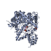 7e9uSC  7e9xC S: Starting model for refinement C: citing same article ( |
|---|---|
| Similar structure data |


-Links
 PDBj
PDBj
- Assembly
| Deposited unit | 
| ||||||||||
|---|---|---|---|---|---|---|---|---|---|---|---|
| 1 | 
| ||||||||||
| 2 | 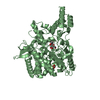
| ||||||||||
| Unit cell |
|
-Components
| #1: Protein | Mass: 65022.859 Da / Num. of mol.: 2 Source method: isolated from a genetically manipulated source Details: A predicted N-terminal transmembrane region (Met1-Leu63) was eliminated. Source: (gene. exp.)  #2: Polysaccharide | #3: Chemical |   Mass: 92.094 Da / Num. of mol.: 3 / Source method: obtained synthetically / Formula: C3H8O3 Mass: 92.094 Da / Num. of mol.: 3 / Source method: obtained synthetically / Formula: C3H8O3#4: Water | ChemComp-HOH / |  Mass: 18.015 Da / Num. of mol.: 841 / Source method: isolated from a natural source / Formula: H2O Mass: 18.015 Da / Num. of mol.: 841 / Source method: isolated from a natural source / Formula: H2OHas ligand of interest | Y | |
|---|
-Experimental details
-Experiment
| Experiment | Method: X-RAY DIFFRACTION / Number of used crystals: 1 |
|---|
- Sample preparation
| Crystal | Density Matthews: 2.52 Å3/Da / Density % sol: 51.13 % |
|---|---|
| Crystal grow | Temperature: 293 K / Method: vapor diffusion, sitting drop Details: 10 mM trehalose, 0.1 M sodium fluoride and 20% PEG3400 |
-Data collection
| Diffraction | Mean temperature: 100 K / Serial crystal experiment: N |
|---|---|
| Diffraction source | Source: SYNCHROTRON / Site: SPring-8 / Beamline: BL41XU / Wavelength: 1 Å |
| Detector | Type: DECTRIS EIGER X 16M / Detector: PIXEL / Date: Oct 6, 2018 |
| Radiation | Protocol: SINGLE WAVELENGTH / Monochromatic (M) / Laue (L): M / Scattering type: x-ray |
| Radiation wavelength | Wavelength: 1 Å / Relative weight: 1 |
| Reflection | Resolution: 1.8→48.16 Å / Num. obs: 122983 / % possible obs: 98.07 % / Redundancy: 9.7 % / Biso Wilson estimate: 21.13 Å2 / CC1/2: 0.995 / Net I/σ(I): 14.45 |
| Reflection shell | Resolution: 1.8→1.864 Å / Num. unique obs: 19210 / CC1/2: 0.924 |
- Processing
| Software |
| |||||||||||||||||||||||||||||||||||||||||||||||||||||||||||||||||||||||||||||||||||||||||||||||||||||||||
|---|---|---|---|---|---|---|---|---|---|---|---|---|---|---|---|---|---|---|---|---|---|---|---|---|---|---|---|---|---|---|---|---|---|---|---|---|---|---|---|---|---|---|---|---|---|---|---|---|---|---|---|---|---|---|---|---|---|---|---|---|---|---|---|---|---|---|---|---|---|---|---|---|---|---|---|---|---|---|---|---|---|---|---|---|---|---|---|---|---|---|---|---|---|---|---|---|---|---|---|---|---|---|---|---|---|---|
| Refinement | Method to determine structure: MOLECULAR REPLACEMENT Starting model: 7e9u Resolution: 1.8→48.16 Å / SU ML: 0.225 / Cross valid method: FREE R-VALUE / σ(F): 1.37 / Phase error: 29.4343 / Stereochemistry target values: GeoStd + Monomer Library
| |||||||||||||||||||||||||||||||||||||||||||||||||||||||||||||||||||||||||||||||||||||||||||||||||||||||||
| Solvent computation | Shrinkage radii: 0.9 Å / VDW probe radii: 1.11 Å / Solvent model: FLAT BULK SOLVENT MODEL | |||||||||||||||||||||||||||||||||||||||||||||||||||||||||||||||||||||||||||||||||||||||||||||||||||||||||
| Displacement parameters | Biso mean: 25.21 Å2 | |||||||||||||||||||||||||||||||||||||||||||||||||||||||||||||||||||||||||||||||||||||||||||||||||||||||||
| Refinement step | Cycle: LAST / Resolution: 1.8→48.16 Å
| |||||||||||||||||||||||||||||||||||||||||||||||||||||||||||||||||||||||||||||||||||||||||||||||||||||||||
| Refine LS restraints |
| |||||||||||||||||||||||||||||||||||||||||||||||||||||||||||||||||||||||||||||||||||||||||||||||||||||||||
| LS refinement shell |
|
