 Movie
Movie Controller

[English] 日本語
 Yorodumi
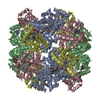
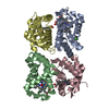
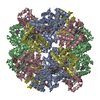
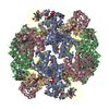
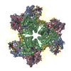
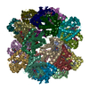
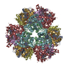
- PDB-7e97: Oxy-deoxy intermediate of 400 kDa giant hemoglobin at 58% oxygen ... -
+ Open data

- Basic information
| Entry | Database: PDB / ID: 7.0E+97 | |||||||||
|---|---|---|---|---|---|---|---|---|---|---|
| Title | Oxy-deoxy intermediate of 400 kDa giant hemoglobin at 58% oxygen saturation | |||||||||
 Components Components |
| |||||||||
 Keywords Keywords | OXYGEN TRANSPORT / allostery / structural change / microspectrometry / crystal processing. | |||||||||
| Function / homology |  Function and homology information Function and homology informationhemoglobin complex / oxygen carrier activity / oxygen binding / iron ion binding / heme binding / extracellular region / metal ion binding Similarity search - Function | |||||||||
| Biological species |  Oligobrachia mashikoi (invertebrata) Oligobrachia mashikoi (invertebrata) | |||||||||
| Method |  X-RAY DIFFRACTION / SYNCHROTRON / MOLECULAR REPLACEMENT / Resolution: 2.7 Å X-RAY DIFFRACTION / SYNCHROTRON / MOLECULAR REPLACEMENT / Resolution: 2.7 Å | |||||||||
 Authors Authors | Numoto, N. / Kawano, Y. / Okumura, H. / Baba, S. / Fukumori, Y. / Miki, K. / Ito, N. | |||||||||
| Funding support |  Japan, 2items Japan, 2items
| |||||||||
 Citation Citation | Journal: Iucrj / Year: 2021 Title: Coarse snapshots of oxygen-dissociation intermediates of a giant hemoglobin elucidated by determining the oxygen saturation in individual subunits in the crystalline state. Authors: Numoto, N. / Kawano, Y. / Okumura, H. / Baba, S. / Fukumori, Y. / Miki, K. / Ito, N. | |||||||||
| History |
|
- Structure visualization
| Structure viewer | Molecule: MolmilJmol/JSmol |
|---|
- Downloads & links
-Download
| PDBx/mmCIF format | 7e97.cif.gz | 124.9 KB | Display | PDBx/mmCIF format |
|---|---|---|---|---|
| PDB format | pdb7e97.ent.gz | 94.6 KB | Display | PDB format |
| PDBx/mmJSON format | 7e97.json.gz | Tree view | PDBx/mmJSON format | |
| Others |  Other downloads Other downloads |
-Validation report
| Arichive directory | https://data.pdbj.org/pub/pdb/validation_reports/e9/7e97ftp://data.pdbj.org/pub/pdb/validation_reports/e9/7e97 | HTTPS FTP |
|---|
-Related structure data
| Related structure data |  7e96C 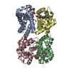 7e98C 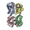 7e99C 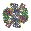 2zfoS S: Starting model for refinement C: citing same article ( |
|---|---|
| Similar structure data |


-Links
 PDBj
PDBj


- Assembly
| Deposited unit | 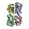
| ||||||||||||
|---|---|---|---|---|---|---|---|---|---|---|---|---|---|
| 1 | x 6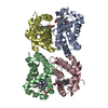
| ||||||||||||
| Unit cell |
| ||||||||||||
| Components on special symmetry positions |
|
-Components
-Extracellular giant hemoglobin major globin subunit ... , 3 types, 3 molecules ABC
| #1: Protein | Mass: 15188.894 Da / Num. of mol.: 1 / Source method: isolated from a natural source / Source: (natural) Oligobrachia mashikoi (invertebrata) / References: UniProt: Q7M419 |
|---|---|
| #2: Protein | Mass: 15327.087 Da / Num. of mol.: 1 / Source method: isolated from a natural source / Source: (natural) Oligobrachia mashikoi (invertebrata) / References: UniProt: Q7M413 |
| #3: Protein | Mass: 15621.285 Da / Num. of mol.: 1 / Source method: isolated from a natural source / Source: (natural) Oligobrachia mashikoi (invertebrata) / References: UniProt: Q7M418 |
-Protein , 1 types, 1 molecules D
| #4: Protein | Mass: 14693.341 Da / Num. of mol.: 1 / Source method: isolated from a natural source / Source: (natural) Oligobrachia mashikoi (invertebrata) / References: UniProt: B1Q3G1 |
|---|
-Non-polymers , 3 types, 25 molecules 


| #5: Chemical | ChemComp-HEM /  Mass: 616.487 Da / Num. of mol.: 4 / Source method: obtained synthetically / Formula: C34H32FeN4O4 / Feature type: SUBJECT OF INVESTIGATION Mass: 616.487 Da / Num. of mol.: 4 / Source method: obtained synthetically / Formula: C34H32FeN4O4 / Feature type: SUBJECT OF INVESTIGATION#6: Chemical | ChemComp-OXY /  Mass: 31.999 Da / Num. of mol.: 4 / Source method: obtained synthetically / Formula: O2 / Feature type: SUBJECT OF INVESTIGATION Mass: 31.999 Da / Num. of mol.: 4 / Source method: obtained synthetically / Formula: O2 / Feature type: SUBJECT OF INVESTIGATION#7: Water | ChemComp-HOH / | Mass: 18.015 Da / Num. of mol.: 17 / Source method: isolated from a natural source / Formula: H2O |
|---|
-Details
| Has ligand of interest | Y |
|---|---|
| Has protein modification | Y |
-Experimental details
-Experiment
| Experiment | Method: X-RAY DIFFRACTION / Number of used crystals: 1 |
|---|
- Sample preparation
| Crystal | Density Matthews: 2.66 Å3/Da / Density % sol: 53.7 % |
|---|---|
| Crystal grow | Temperature: 293 K / Method: vapor diffusion, sitting drop / pH: 8 Details: 11-12 % (w/v) polyethylene glycol (PEG) 10,000, 10 mM CaCl2, 200 mM Tris-HCl pH 8.0 |
-Data collection
| Diffraction | Mean temperature: 95 K / Serial crystal experiment: N |
|---|---|
| Diffraction source | Source: SYNCHROTRON / Site: Photon Factory / Beamline: BL-17A / Wavelength: 0.98 Å |
| Detector | Type: DECTRIS PILATUS3 6M / Detector: PIXEL / Date: Jun 28, 2016 |
| Radiation | Protocol: SINGLE WAVELENGTH / Monochromatic (M) / Laue (L): M / Scattering type: x-ray |
| Radiation wavelength | Wavelength: 0.98 Å / Relative weight: 1 |
| Reflection | Resolution: 2.7→50 Å / Num. obs: 18081 / % possible obs: 99.8 % / Redundancy: 9.7 % / Biso Wilson estimate: 59.17 Å2 / CC1/2: 0.998 / Rsym value: 0.208 / Net I/σ(I): 9 |
| Reflection shell | Resolution: 2.7→2.87 Å / Redundancy: 9.4 % / Mean I/σ(I) obs: 1.5 / Num. unique obs: 2846 / CC1/2: 0.583 / Rsym value: 1.37 / % possible all: 99.2 |
- Processing
| Software |
| |||||||||||||||||||||||||||||||||||||||||||||||||
|---|---|---|---|---|---|---|---|---|---|---|---|---|---|---|---|---|---|---|---|---|---|---|---|---|---|---|---|---|---|---|---|---|---|---|---|---|---|---|---|---|---|---|---|---|---|---|---|---|---|---|
| Refinement | Method to determine structure: MOLECULAR REPLACEMENT Starting model: 2ZFO Resolution: 2.7→47.4 Å / SU ML: 0.3498 / Cross valid method: FREE R-VALUE / σ(F): 1.35 / Phase error: 28.0503 / Stereochemistry target values: GeoStd + Monomer Library
| |||||||||||||||||||||||||||||||||||||||||||||||||
| Solvent computation | Shrinkage radii: 0.9 Å / VDW probe radii: 1.11 Å / Solvent model: FLAT BULK SOLVENT MODEL | |||||||||||||||||||||||||||||||||||||||||||||||||
| Displacement parameters | Biso mean: 61.18 Å2 | |||||||||||||||||||||||||||||||||||||||||||||||||
| Refinement step | Cycle: LAST / Resolution: 2.7→47.4 Å
| |||||||||||||||||||||||||||||||||||||||||||||||||
| Refine LS restraints |
| |||||||||||||||||||||||||||||||||||||||||||||||||
| LS refinement shell |
|
