 Movie
Movie Controller

[English] 日本語
 Yorodumi
- PDB-7e8p: Crystal structure of a Flavin-dependent Monooxygenase HadA wild t... -
+ Open data

- Basic information
| Entry | Database: PDB / ID: 7e8p | |||||||||
|---|---|---|---|---|---|---|---|---|---|---|
| Title | Crystal structure of a Flavin-dependent Monooxygenase HadA wild type complexed with reduced FAD and 4-nitrophenol | |||||||||
 Components Components | Chlorophenol monooxygenase | |||||||||
 Keywords Keywords | OXIDOREDUCTASE / flavin monooxygenase / chlorophenol 4-monooxygenase | |||||||||
| Function / homology |  Function and homology information Function and homology informationoxidoreductase activity, acting on the CH-CH group of donors / monooxygenase activity Similarity search - Function | |||||||||
| Biological species |  Ralstonia pickettii DTP0602 (bacteria) Ralstonia pickettii DTP0602 (bacteria) | |||||||||
| Method |  X-RAY DIFFRACTION / MOLECULAR REPLACEMENT / Resolution: 2.3 Å X-RAY DIFFRACTION / MOLECULAR REPLACEMENT / Resolution: 2.3 Å | |||||||||
| Model details | Crystal structure of Flavin-dependent Monooxygenase HadA with FADH2 and 4-nitophenol | |||||||||
 Authors Authors | Pimviriyakul, P. / Jaruwat, A. / Chitnumsub, P. / Chaiyen, P. | |||||||||
| Funding support |  Thailand, 2items Thailand, 2items
| |||||||||
 Citation Citation | Journal: J.Biol.Chem. / Year: 2021 Title: Structural insights into a flavin-dependent dehalogenase HadA explain catalysis and substrate inhibition via quadruple pi-stacking. Authors: Pimviriyakul, P. / Jaruwat, A. / Chitnumsub, P. / Chaiyen, P. | |||||||||
| History |
|
- Structure visualization
| Structure viewer | Molecule: MolmilJmol/JSmol |
|---|
- Downloads & links
-Download
| PDBx/mmCIF format | 7e8p.cif.gz | 411 KB | Display | PDBx/mmCIF format |
|---|---|---|---|---|
| PDB format | pdb7e8p.ent.gz | 332.7 KB | Display | PDB format |
| PDBx/mmJSON format | 7e8p.json.gz | Tree view | PDBx/mmJSON format | |
| Others |  Other downloads Other downloads |
-Validation report
| Arichive directory | https://data.pdbj.org/pub/pdb/validation_reports/e8/7e8pftp://data.pdbj.org/pub/pdb/validation_reports/e8/7e8p | HTTPS FTP |
|---|
-Related structure data
| Related structure data |  7e8qC  6jhmS S: Starting model for refinement C: citing same article ( |
|---|---|
| Similar structure data |


-Links
 PDBj
PDBj
- Assembly
| Deposited unit | 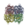
| ||||||||
|---|---|---|---|---|---|---|---|---|---|
| 1 |
| ||||||||
| Unit cell |
|
-Components
| #1: Protein | Mass: 58623.164 Da / Num. of mol.: 4 / Fragment: RESIDUES 1-517 Source method: isolated from a genetically manipulated source Source: (gene. exp.) Ralstonia pickettii DTP0602 (bacteria) / Gene: hadA / Plasmid: pET11a / Production host: References: UniProt: Q53008, 4-hydroxyphenylacetate 3-monooxygenase #2: Chemical | ChemComp-FDA /   Mass: 787.566 Da / Num. of mol.: 4 / Source method: obtained synthetically / Formula: C27H35N9O15P2 / Feature type: SUBJECT OF INVESTIGATION Mass: 787.566 Da / Num. of mol.: 4 / Source method: obtained synthetically / Formula: C27H35N9O15P2 / Feature type: SUBJECT OF INVESTIGATION#3: Chemical |   Mass: 139.109 Da / Num. of mol.: 3 / Source method: obtained synthetically / Formula: C6H5NO3 Mass: 139.109 Da / Num. of mol.: 3 / Source method: obtained synthetically / Formula: C6H5NO3#4: Water | ChemComp-HOH / |  Mass: 18.015 Da / Num. of mol.: 878 / Source method: isolated from a natural source / Formula: H2O Mass: 18.015 Da / Num. of mol.: 878 / Source method: isolated from a natural source / Formula: H2OHas ligand of interest | Y | |
|---|
-Experimental details
-Experiment
| Experiment | Method: X-RAY DIFFRACTION / Number of used crystals: 1 |
|---|
- Sample preparation
| Crystal | Density Matthews: 2.85 Å3/Da / Density % sol: 56.79 % / Mosaicity: 0 ° |
|---|---|
| Crystal grow | Temperature: 298 K / Method: microbatch / pH: 6.5 Details: 0.1 M Bis-Tris propane pH 6.5, 0.35 M sodium citrate tribasic dihydrate and 24% w/v PEG3350 |
-Data collection
| Diffraction | Mean temperature: 100 K / Serial crystal experiment: N | ||||||||||||||||||||||||||||||
|---|---|---|---|---|---|---|---|---|---|---|---|---|---|---|---|---|---|---|---|---|---|---|---|---|---|---|---|---|---|---|---|
| Diffraction source | Source: ROTATING ANODE / Type: BRUKER TURBO X-RAY SOURCE / Wavelength: 1.54 Å | ||||||||||||||||||||||||||||||
| Detector | Type: BRUKER PHOTON 100 / Detector: CMOS / Date: Mar 13, 2018 | ||||||||||||||||||||||||||||||
| Radiation | Protocol: SINGLE WAVELENGTH / Monochromatic (M) / Laue (L): M / Scattering type: x-ray | ||||||||||||||||||||||||||||||
| Radiation wavelength | Wavelength: 1.54 Å / Relative weight: 1 | ||||||||||||||||||||||||||||||
| Reflection | Resolution: 2.3→20.74 Å / Num. obs: 118305 / % possible obs: 99.3 % / Redundancy: 5 % / CC1/2: 0.993 / Rmerge(I) obs: 0.11 / Rpim(I) all: 0.052 / Rrim(I) all: 0.122 / Net I/σ(I): 9.9 / Num. measured all: 586952 | ||||||||||||||||||||||||||||||
| Reflection shell | Diffraction-ID: 1
|
- Processing
| Software |
| ||||||||||||||||||||||||||||||||||||||||||||||||||||||||||||
|---|---|---|---|---|---|---|---|---|---|---|---|---|---|---|---|---|---|---|---|---|---|---|---|---|---|---|---|---|---|---|---|---|---|---|---|---|---|---|---|---|---|---|---|---|---|---|---|---|---|---|---|---|---|---|---|---|---|---|---|---|---|
| Refinement | Method to determine structure: MOLECULAR REPLACEMENT Starting model: 6JHM Resolution: 2.3→20.71 Å / Cor.coef. Fo:Fc: 0.922 / Cor.coef. Fo:Fc free: 0.871 / SU B: 8.258 / SU ML: 0.194 / Cross valid method: THROUGHOUT / σ(F): 0 / ESU R: 0.306 / ESU R Free: 0.243 / Stereochemistry target values: MAXIMUM LIKELIHOOD Details: HYDROGENS HAVE BEEN ADDED IN THE RIDING POSITIONS U VALUES : REFINED INDIVIDUALLY
| ||||||||||||||||||||||||||||||||||||||||||||||||||||||||||||
| Solvent computation | Ion probe radii: 0.8 Å / Shrinkage radii: 0.8 Å / VDW probe radii: 1.4 Å / Solvent model: MASK | ||||||||||||||||||||||||||||||||||||||||||||||||||||||||||||
| Displacement parameters | Biso max: 124.52 Å2 / Biso mean: 20.662 Å2 / Biso min: 2.02 Å2
| ||||||||||||||||||||||||||||||||||||||||||||||||||||||||||||
| Refinement step | Cycle: final / Resolution: 2.3→20.71 Å
| ||||||||||||||||||||||||||||||||||||||||||||||||||||||||||||
| Refine LS restraints |
| ||||||||||||||||||||||||||||||||||||||||||||||||||||||||||||
| LS refinement shell | Resolution: 2.3→2.359 Å / Rfactor Rfree error: 0 / Total num. of bins used: 20
|
