 Movie
Movie Controller

[English] 日本語
 Yorodumi
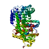
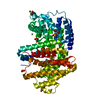
- PDB-7e6w: Crystal structure of Sesquisabinene B Synthase 1 mutant G418A and... -
+ Open data

- Basic information
| Entry | Database: PDB / ID: 7e6w | ||||||
|---|---|---|---|---|---|---|---|
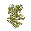
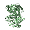
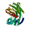
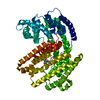
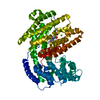
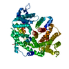
| Title | Crystal structure of Sesquisabinene B Synthase 1 mutant G418A and F419N | ||||||
 Components Components | Sesquisabinene B synthase 1 | ||||||
 Keywords Keywords | LYASE / Terpene synthase / Santalum album / Farnesyl Pyrophosphate / sandalwood oil | ||||||
| Function / homology |  Function and homology information Function and homology informationditerpenoid biosynthetic process / terpene synthase activity / magnesium ion binding Similarity search - Function | ||||||
| Biological species |  Santalum album (white sandalwood) Santalum album (white sandalwood) | ||||||
| Method |  X-RAY DIFFRACTION / SYNCHROTRON / MOLECULAR REPLACEMENT / Resolution: 3.1 Å X-RAY DIFFRACTION / SYNCHROTRON / MOLECULAR REPLACEMENT / Resolution: 3.1 Å | ||||||
 Authors Authors | Singh, S. / Thulasiram, H.V. / Kulkarni, K.A. | ||||||
| Funding support |  India, 1items India, 1items
| ||||||
 Citation Citation | Journal: To Be Published Title: Crystal structure of Sesquisabinene B Synthase 1 mutant G418A and F419N Authors: Singh, S. / Thulasiram, H.V. / Kulkarni, K.A. | ||||||
| History |
|
- Structure visualization
| Structure viewer | Molecule: MolmilJmol/JSmol |
|---|
- Downloads & links
-Download
| PDBx/mmCIF format | 7e6w.cif.gz | 424.5 KB | Display | PDBx/mmCIF format |
|---|---|---|---|---|
| PDB format | pdb7e6w.ent.gz | 351.4 KB | Display | PDB format |
| PDBx/mmJSON format | 7e6w.json.gz | Tree view | PDBx/mmJSON format | |
| Others |  Other downloads Other downloads |
-Validation report
| Summary document | 7e6w_validation.pdf.gz | 454.1 KB | Display | wwPDB validaton report |
|---|---|---|---|---|
| Full document | 7e6w_full_validation.pdf.gz | 480.3 KB | Display | |
| Data in XML | 7e6w_validation.xml.gz | 37 KB | Display | |
| Data in CIF | 7e6w_validation.cif.gz | 50.4 KB | Display | |
| Arichive directory | https://data.pdbj.org/pub/pdb/validation_reports/e6/7e6wftp://data.pdbj.org/pub/pdb/validation_reports/e6/7e6w | HTTPS FTP |
-Related structure data
| Related structure data |  6k16S S: Starting model for refinement |
|---|---|
| Similar structure data |
-Links
 PDBj
PDBj

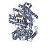
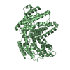
- Assembly
| Deposited unit | 
| ||||||||
|---|---|---|---|---|---|---|---|---|---|
| 1 | 
| ||||||||
| 2 | 
| ||||||||
| Unit cell |
|
-Components
| #1: Protein | Mass: 64248.648 Da / Num. of mol.: 2 / Mutation: G418A, F419N Source method: isolated from a genetically manipulated source Source: (gene. exp.) Santalum album (white sandalwood) / Plasmid: pOPINSS / Production host:  |
|---|
-Experimental details
-Experiment
| Experiment | Method: X-RAY DIFFRACTION / Number of used crystals: 1 |
|---|
- Sample preparation
| Crystal | Density Matthews: 2.39 Å3/Da / Density % sol: 48.55 % |
|---|---|
| Crystal grow | Temperature: 293.15 K / Method: vapor diffusion, sitting drop / pH: 6.5 Details: 0.1M MES (pH-6.5), 0.1M Magnesium Acetate, 10% PEG 10,000 |
-Data collection
| Diffraction | Mean temperature: 100 K / Serial crystal experiment: N |
|---|---|
| Diffraction source | Source: SYNCHROTRON / Site: Diamond  / Beamline: I03 / Wavelength: 0.976 Å / Beamline: I03 / Wavelength: 0.976 Å |
| Detector | Type: DECTRIS EIGER X 16M / Detector: PIXEL / Date: Jan 27, 2020 |
| Radiation | Protocol: SINGLE WAVELENGTH / Monochromatic (M) / Laue (L): M / Scattering type: x-ray |
| Radiation wavelength | Wavelength: 0.976 Å / Relative weight: 1 |
| Reflection | Resolution: 3.1→47.58 Å / Num. obs: 21943 / % possible obs: 99.8 % / Redundancy: 6.2 % / Biso Wilson estimate: 86.54 Å2 / CC1/2: 0.999 / Rmerge(I) obs: 0.081 / Rpim(I) all: 0.053 / Rrim(I) all: 0.097 / Net I/σ(I): 11.5 |
| Reflection shell | Resolution: 3.1→3.31 Å / Redundancy: 6.4 % / Rmerge(I) obs: 0.859 / Mean I/σ(I) obs: 1.7 / Num. unique obs: 3923 / CC1/2: 0.895 / Rpim(I) all: 0.554 / Rrim(I) all: 1 / % possible all: 99.8 |
- Processing
| Software |
| |||||||||||||||||||||||||||||||||||||||||||||||||||||||||||||||||||||||||||
|---|---|---|---|---|---|---|---|---|---|---|---|---|---|---|---|---|---|---|---|---|---|---|---|---|---|---|---|---|---|---|---|---|---|---|---|---|---|---|---|---|---|---|---|---|---|---|---|---|---|---|---|---|---|---|---|---|---|---|---|---|---|---|---|---|---|---|---|---|---|---|---|---|---|---|---|---|
| Refinement | Method to determine structure: MOLECULAR REPLACEMENT Starting model: 6K16 Resolution: 3.1→45.61 Å / SU ML: 0.54 / Cross valid method: FREE R-VALUE / σ(F): 1.97 / Phase error: 34.68 / Stereochemistry target values: ML
| |||||||||||||||||||||||||||||||||||||||||||||||||||||||||||||||||||||||||||
| Solvent computation | Shrinkage radii: 0.9 Å / VDW probe radii: 1.11 Å / Solvent model: FLAT BULK SOLVENT MODEL | |||||||||||||||||||||||||||||||||||||||||||||||||||||||||||||||||||||||||||
| Refinement step | Cycle: LAST / Resolution: 3.1→45.61 Å
| |||||||||||||||||||||||||||||||||||||||||||||||||||||||||||||||||||||||||||
| Refine LS restraints |
| |||||||||||||||||||||||||||||||||||||||||||||||||||||||||||||||||||||||||||
| LS refinement shell |
| |||||||||||||||||||||||||||||||||||||||||||||||||||||||||||||||||||||||||||
| Refinement TLS params. | Method: refined / Refine-ID: X-RAY DIFFRACTION
| |||||||||||||||||||||||||||||||||||||||||||||||||||||||||||||||||||||||||||
| Refinement TLS group |
|
