 Movie
Movie Controller

[English] 日本語
 Yorodumi
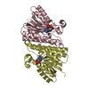
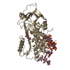
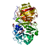
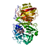
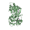
- PDB-7e3x: Crystal structure of SDR family NAD(P)-dependent oxidoreductase f... -
+ Open data

- Basic information
| Entry | Database: PDB / ID: 7e3x | ||||||
|---|---|---|---|---|---|---|---|
| Title | Crystal structure of SDR family NAD(P)-dependent oxidoreductase from exiguobacterium | ||||||
 Components Components | Oxidoreductase | ||||||
 Keywords Keywords | OXIDOREDUCTASE / mutant / short chain reductase | ||||||
| Function / homology |  Function and homology information Function and homology information | ||||||
| Biological species |  Exiguobacterium sp. KKBO11 (bacteria) Exiguobacterium sp. KKBO11 (bacteria) | ||||||
| Method |  X-RAY DIFFRACTION / SYNCHROTRON / MOLECULAR REPLACEMENT / Resolution: 2.58 Å X-RAY DIFFRACTION / SYNCHROTRON / MOLECULAR REPLACEMENT / Resolution: 2.58 Å | ||||||
 Authors Authors | Chen, L. / Tang, J. / Yuan, S. / Zhang, F. / Chen, S. | ||||||
 Citation Citation | Journal: Catalysis Science And Technology / Year: 2021 Title: Structure-guided evolution of a ketoreductase forefficient and stereoselective bioreduction of bulkyalpha-aminobeta-keto esters Authors: Tang, J. / Chen, L. / Zhang, L. / Ni, G. / Yu, J. / Wang, H. / Zhang, F. / Yuan, S. / Feng, M. / Che, S. | ||||||
| History |
|
- Structure visualization
| Structure viewer | Molecule: MolmilJmol/JSmol |
|---|
- Downloads & links
-Download
| PDBx/mmCIF format | 7e3x.cif.gz | 110.8 KB | Display | PDBx/mmCIF format |
|---|---|---|---|---|
| PDB format | pdb7e3x.ent.gz | 83.5 KB | Display | PDB format |
| PDBx/mmJSON format | 7e3x.json.gz | Tree view | PDBx/mmJSON format | |
| Others |  Other downloads Other downloads |
-Validation report
| Arichive directory | https://data.pdbj.org/pub/pdb/validation_reports/e3/7e3xftp://data.pdbj.org/pub/pdb/validation_reports/e3/7e3x | HTTPS FTP |
|---|
-Related structure data
| Related structure data |  7e24C  7e28C  5t2uS S: Starting model for refinement C: citing same article ( |
|---|---|
| Similar structure data |


-Links
 PDBj
PDBj


- Assembly
| Deposited unit | 
| ||||||||||
|---|---|---|---|---|---|---|---|---|---|---|---|
| 1 |
| ||||||||||
| Unit cell |
|
-Components
| #1: Protein | Mass: 28159.857 Da / Num. of mol.: 2 / Mutation: F88V, V127I, A138L, R142M, A190V, S193A Source method: isolated from a genetically manipulated source Source: (gene. exp.) Exiguobacterium sp. KKBO11 (bacteria) / Gene: AYO36_14830 / Production host: #2: Chemical |   Mass: 743.405 Da / Num. of mol.: 2 / Source method: obtained synthetically / Formula: C21H28N7O17P3 Mass: 743.405 Da / Num. of mol.: 2 / Source method: obtained synthetically / Formula: C21H28N7O17P3#3: Water | ChemComp-HOH / |  Mass: 18.015 Da / Num. of mol.: 48 / Source method: isolated from a natural source / Formula: H2O Mass: 18.015 Da / Num. of mol.: 48 / Source method: isolated from a natural source / Formula: H2OHas ligand of interest | N | |
|---|
-Experimental details
-Experiment
| Experiment | Method: X-RAY DIFFRACTION / Number of used crystals: 1 |
|---|
- Sample preparation
| Crystal | Density Matthews: 2.49 Å3/Da / Density % sol: 50.62 % |
|---|---|
| Crystal grow | Temperature: 293.15 K / Method: vapor diffusion, hanging drop / Details: PEG 3350 |
-Data collection
| Diffraction | Mean temperature: 100 K / Serial crystal experiment: N |
|---|---|
| Diffraction source | Source: SYNCHROTRON / Site: SSRF  / Beamline: BL18U1 / Wavelength: 0.979 Å / Beamline: BL18U1 / Wavelength: 0.979 Å |
| Detector | Type: MAR CCD 130 mm / Detector: CCD / Date: Dec 25, 2020 |
| Radiation | Protocol: SINGLE WAVELENGTH / Monochromatic (M) / Laue (L): M / Scattering type: x-ray |
| Radiation wavelength | Wavelength: 0.979 Å / Relative weight: 1 |
| Reflection | Resolution: 2.58→50 Å / Num. obs: 17891 / % possible obs: 97.7 % / Redundancy: 4.8 % / Biso Wilson estimate: 29.57 Å2 / CC1/2: 0.948 / Net I/σ(I): 1.6 |
| Reflection shell | Resolution: 2.6→2.64 Å / Mean I/σ(I) obs: 1.4 / Num. unique obs: 821 / CC1/2: 0.57 |
- Processing
| Software |
| ||||||||||||||||
|---|---|---|---|---|---|---|---|---|---|---|---|---|---|---|---|---|---|
| Refinement | Method to determine structure: MOLECULAR REPLACEMENT Starting model: 5t2u Resolution: 2.58→44.76 Å / Cross valid method: THROUGHOUT
| ||||||||||||||||
| Displacement parameters | Biso max: 71.02 Å2 / Biso mean: 28.0926 Å2 / Biso min: 12.48 Å2 | ||||||||||||||||
| Refinement step | Cycle: LAST / Resolution: 2.58→44.76 Å
| ||||||||||||||||
| LS refinement shell | Resolution: 2.58→2.6693 Å
|
