 Movie
Movie Controller

[English] 日本語
 Yorodumi
- PDB-7dyg: Histone lysine demethylase 4D (KDM4D) in complex with the inhibit... -
+ Open data

- Basic information
| Entry | Database: PDB / ID: 7dyg | ||||||
|---|---|---|---|---|---|---|---|
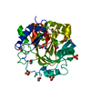
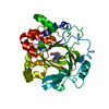
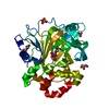
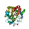
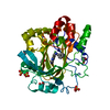
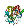
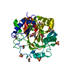
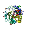
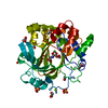
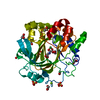
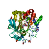
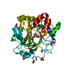
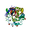
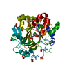
| Title | Histone lysine demethylase 4D (KDM4D) in complex with the inhibitor 2-(1H-pyrazol-3-yl)isonicotinic acid | ||||||
 Components Components | Lysine-specific demethylase 4D | ||||||
 Keywords Keywords | OXIDOREDUCTASE / Complex / Inhibitor | ||||||
| Function / homology |  Function and homology information Function and homology informationpositive regulation of chromatin binding / [histone H3]-trimethyl-L-lysine9 demethylase / histone H3K9me2/H3K9me3 demethylase activity / positive regulation of double-strand break repair via nonhomologous end joining / regulation of protein phosphorylation / histone H3K9 demethylase activity / histone demethylase activity / pericentric heterochromatin / cellular response to ionizing radiation / HDMs demethylate histones ...positive regulation of chromatin binding / [histone H3]-trimethyl-L-lysine9 demethylase / histone H3K9me2/H3K9me3 demethylase activity / positive regulation of double-strand break repair via nonhomologous end joining / regulation of protein phosphorylation / histone H3K9 demethylase activity / histone demethylase activity / pericentric heterochromatin / cellular response to ionizing radiation / HDMs demethylate histones / chromatin DNA binding / double-strand break repair via homologous recombination / site of double-strand break / regulation of gene expression / blood microparticle / damaged DNA binding / chromatin remodeling / inflammatory response / chromatin / nucleoplasm / metal ion binding / nucleus Similarity search - Function | ||||||
| Biological species |  Homo sapiens (human) Homo sapiens (human) | ||||||
| Method |  X-RAY DIFFRACTION / SYNCHROTRON / MOLECULAR REPLACEMENT / Resolution: 2 Å X-RAY DIFFRACTION / SYNCHROTRON / MOLECULAR REPLACEMENT / Resolution: 2 Å | ||||||
 Authors Authors | Wang, T. / Yang, L. | ||||||
 Citation Citation | Journal: To Be Published Title: Crystal structure of histone lysine demethylase 4D (KDM4D) in complex with the inhibitor 2-(1H-pyrazol-3-yl)isonicotinic acid Authors: Wang, T. / Yang, L. | ||||||
| History |
|
- Structure visualization
| Structure viewer | Molecule: MolmilJmol/JSmol |
|---|
- Downloads & links
-Download
| PDBx/mmCIF format | 7dyg.cif.gz | 145 KB | Display | PDBx/mmCIF format |
|---|---|---|---|---|
| PDB format | pdb7dyg.ent.gz | 113 KB | Display | PDB format |
| PDBx/mmJSON format | 7dyg.json.gz | Tree view | PDBx/mmJSON format | |
| Others |  Other downloads Other downloads |
-Validation report
| Arichive directory | https://data.pdbj.org/pub/pdb/validation_reports/dy/7dygftp://data.pdbj.org/pub/pdb/validation_reports/dy/7dyg | HTTPS FTP |
|---|
-Related structure data
| Related structure data | 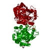 4d6qS S: Starting model for refinement |
|---|---|
| Similar structure data |


-Links
 PDBj
PDBj
- Assembly
| Deposited unit | 
| ||||||||
|---|---|---|---|---|---|---|---|---|---|
| 1 |
| ||||||||
| Unit cell |
| ||||||||
| Components on special symmetry positions |
|
-Components
| #1: Protein | Mass: 38093.156 Da / Num. of mol.: 1 Source method: isolated from a genetically manipulated source Source: (gene. exp.) Homo sapiens (human) / Gene: KDM4D, JHDM3D, JMJD2D / Production host:  References: UniProt: Q6B0I6, [histone H3]-trimethyl-L-lysine9 demethylase |
|---|---|
| #2: Chemical | ChemComp-0WS /   Mass: 189.171 Da / Num. of mol.: 1 / Source method: obtained synthetically / Formula: C9H7N3O2 / Feature type: SUBJECT OF INVESTIGATION Mass: 189.171 Da / Num. of mol.: 1 / Source method: obtained synthetically / Formula: C9H7N3O2 / Feature type: SUBJECT OF INVESTIGATION |
| #3: Chemical | ChemComp-FE /   Mass: 55.845 Da / Num. of mol.: 1 / Source method: obtained synthetically / Formula: Fe Mass: 55.845 Da / Num. of mol.: 1 / Source method: obtained synthetically / Formula: Fe |
| #4: Water | ChemComp-HOH /  Mass: 18.015 Da / Num. of mol.: 327 / Source method: isolated from a natural source / Formula: H2O Mass: 18.015 Da / Num. of mol.: 327 / Source method: isolated from a natural source / Formula: H2O |
| Has ligand of interest | Y |
| Has protein modification | Y |
-Experimental details
-Experiment
| Experiment | Method: X-RAY DIFFRACTION / Number of used crystals: 1 |
|---|
- Sample preparation
| Crystal | Density Matthews: 2.55 Å3/Da / Density % sol: 51.82 % |
|---|---|
| Crystal grow | Temperature: 291.15 K / Method: vapor diffusion, sitting drop Details: 0.2M potassium citrate tribasic monohydrate PH 8.3, 20% w/v PEG 3350 |
-Data collection
| Diffraction | Mean temperature: 100 K / Serial crystal experiment: N |
|---|---|
| Diffraction source | Source: SYNCHROTRON / Site: NFPSS  / Beamline: BL19U1 / Wavelength: 0.9793 Å / Beamline: BL19U1 / Wavelength: 0.9793 Å |
| Detector | Type: DECTRIS PILATUS3 X CdTe 1M / Detector: PIXEL / Date: Jan 13, 2020 |
| Radiation | Protocol: SINGLE WAVELENGTH / Monochromatic (M) / Laue (L): M / Scattering type: x-ray |
| Radiation wavelength | Wavelength: 0.9793 Å / Relative weight: 1 |
| Reflection | Resolution: 2→50 Å / Num. obs: 27588 / % possible obs: 100 % / Redundancy: 25.6 % / Rmerge(I) obs: 0.095 / Net I/σ(I): 38.2 |
| Reflection shell | Resolution: 2→2.03 Å / Rmerge(I) obs: 0.238 / Num. unique obs: 1333 |
- Processing
| Software |
| ||||||||||||||||||||||||||||||||||||||||||||||||||||||||||||||||||
|---|---|---|---|---|---|---|---|---|---|---|---|---|---|---|---|---|---|---|---|---|---|---|---|---|---|---|---|---|---|---|---|---|---|---|---|---|---|---|---|---|---|---|---|---|---|---|---|---|---|---|---|---|---|---|---|---|---|---|---|---|---|---|---|---|---|---|---|
| Refinement | Method to determine structure: MOLECULAR REPLACEMENT Starting model: 4d6q Resolution: 2→25.978 Å / SU ML: 0.14 / Cross valid method: THROUGHOUT / σ(F): 1.34 / Phase error: 17.29 / Stereochemistry target values: ML
| ||||||||||||||||||||||||||||||||||||||||||||||||||||||||||||||||||
| Solvent computation | Shrinkage radii: 0.9 Å / VDW probe radii: 1.11 Å / Solvent model: FLAT BULK SOLVENT MODEL | ||||||||||||||||||||||||||||||||||||||||||||||||||||||||||||||||||
| Displacement parameters | Biso max: 100.83 Å2 / Biso mean: 27.7658 Å2 / Biso min: 5.44 Å2 | ||||||||||||||||||||||||||||||||||||||||||||||||||||||||||||||||||
| Refinement step | Cycle: final / Resolution: 2→25.978 Å
| ||||||||||||||||||||||||||||||||||||||||||||||||||||||||||||||||||
| Refine LS restraints |
| ||||||||||||||||||||||||||||||||||||||||||||||||||||||||||||||||||
| LS refinement shell | Refine-ID: X-RAY DIFFRACTION / Rfactor Rfree error: 0
|
