 Movie
Movie Controller


+ Open data

- Basic information
| Entry | Database: PDB / ID: 7dus | ||||||
|---|---|---|---|---|---|---|---|
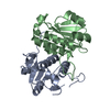
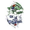
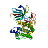
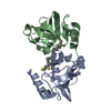
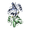
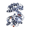
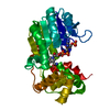
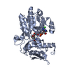
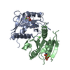
| Title | Crystal structure of Mei2-RRM3 domain in S.pombe | ||||||
 Components Components | Meiosis protein mei2 | ||||||
 Keywords Keywords | RNA BINDING PROTEIN / Meiosis / Mmi / RRM / Pombe | ||||||
| Function / homology |  Function and homology information Function and homology informationTor2-Mei2-Ste11 complex / cell cycle switching, mitotic to meiotic cell cycle / negative regulation of conjugation with zygote / positive regulation of metaphase/anaphase transition of meiosis II / Mei2 nuclear dot complex / positive regulation of meiotic cell cycle / poly(U) RNA binding / nuclear chromosome / lncRNA binding / protein sequestering activity ...Tor2-Mei2-Ste11 complex / cell cycle switching, mitotic to meiotic cell cycle / negative regulation of conjugation with zygote / positive regulation of metaphase/anaphase transition of meiosis II / Mei2 nuclear dot complex / positive regulation of meiotic cell cycle / poly(U) RNA binding / nuclear chromosome / lncRNA binding / protein sequestering activity / meiotic cell cycle / RNA binding / nucleus / cytosol / cytoplasm Similarity search - Function | ||||||
| Biological species |  | ||||||
| Method |  X-RAY DIFFRACTION / SYNCHROTRON / SAD / Resolution: 2.5 Å X-RAY DIFFRACTION / SYNCHROTRON / SAD / Resolution: 2.5 Å | ||||||
 Authors Authors | Wu, B.X. / Xu, J.H. / Ma, J.B. | ||||||
 Citation Citation | Journal: To Be Published Title: Crystal structure of Mei2-RRM3 domain in S.pombe Authors: Wu, B.X. / Xu, J.H. / Ma, J.B. | ||||||
| History |
|
- Structure visualization
| Structure viewer | Molecule: MolmilJmol/JSmol |
|---|
- Downloads & links
-Download
| PDBx/mmCIF format | 7dus.cif.gz | 42.4 KB | Display | PDBx/mmCIF format |
|---|---|---|---|---|
| PDB format | pdb7dus.ent.gz | 27.7 KB | Display | PDB format |
| PDBx/mmJSON format | 7dus.json.gz | Tree view | PDBx/mmJSON format | |
| Others |  Other downloads Other downloads |
-Validation report
| Summary document | 7dus_validation.pdf.gz | 445.3 KB | Display | wwPDB validaton report |
|---|---|---|---|---|
| Full document | 7dus_full_validation.pdf.gz | 446 KB | Display | |
| Data in XML | 7dus_validation.xml.gz | 7.6 KB | Display | |
| Data in CIF | 7dus_validation.cif.gz | 9.2 KB | Display | |
| Arichive directory | https://data.pdbj.org/pub/pdb/validation_reports/du/7dusftp://data.pdbj.org/pub/pdb/validation_reports/du/7dus | HTTPS FTP |
-Related structure data
| Similar structure data |
|---|

-Links
 PDBj
PDBj- Assembly
| Deposited unit | 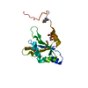
| ||||||||||||
|---|---|---|---|---|---|---|---|---|---|---|---|---|---|
| 1 | 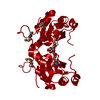
| ||||||||||||
| Unit cell |
|
-Components
| #1: Protein | Mass: 17845.762 Da / Num. of mol.: 1 Source method: isolated from a genetically manipulated source Source: (gene. exp.) Strain: 972 / ATCC 24843 / Gene: mei2, SPAC27D7.03c / Production host:  |
|---|---|
| #2: Chemical | ChemComp-CIT /   Mass: 192.124 Da / Num. of mol.: 1 / Source method: obtained synthetically / Formula: C6H8O7 Mass: 192.124 Da / Num. of mol.: 1 / Source method: obtained synthetically / Formula: C6H8O7 |
| #3: Water | ChemComp-HOH /  Mass: 18.015 Da / Num. of mol.: 17 / Source method: isolated from a natural source / Formula: H2O Mass: 18.015 Da / Num. of mol.: 17 / Source method: isolated from a natural source / Formula: H2O |
| Has ligand of interest | N |
| Has protein modification | Y |
-Experimental details
-Experiment
| Experiment | Method: X-RAY DIFFRACTION / Number of used crystals: 1 |
|---|
- Sample preparation
| Crystal | Density Matthews: 3.99 Å3/Da / Density % sol: 69.2 % |
|---|---|
| Crystal grow | Temperature: 293 K / Method: vapor diffusion, hanging drop / pH: 8.6 / Details: 15% PEG 3350, 0.2MTri-Sodium Citrate Tris pH 8.6 |
-Data collection
| Diffraction | Mean temperature: 100 K / Serial crystal experiment: N |
|---|---|
| Diffraction source | Source: SYNCHROTRON / Site: SSRF  / Beamline: BL19U1 / Wavelength: 0.9793 Å / Beamline: BL19U1 / Wavelength: 0.9793 Å |
| Detector | Type: DECTRIS PILATUS 6M / Detector: PIXEL / Date: Nov 3, 2015 |
| Radiation | Protocol: SINGLE WAVELENGTH / Monochromatic (M) / Laue (L): M / Scattering type: x-ray |
| Radiation wavelength | Wavelength: 0.9793 Å / Relative weight: 1 |
| Reflection | Resolution: 2.5→30 Å / Num. obs: 10649 / % possible obs: 99.9 % / Redundancy: 16.5 % / CC1/2: 0.933 / CC star: 0.98 / Rmerge(I) obs: 0.125 / Rpim(I) all: 0.028 / Rrim(I) all: 0.117 / Net I/σ(I): 21.879 |
| Reflection shell | Resolution: 2.5→2.59 Å / Redundancy: 14.3 % / Mean I/σ(I) obs: 2.16 / Num. unique obs: 1034 / CC1/2: 0.771 / CC star: 0.933 / Rpim(I) all: 0.271 / % possible all: 100 |
- Processing
| Software |
| ||||||||||||||||||||||||
|---|---|---|---|---|---|---|---|---|---|---|---|---|---|---|---|---|---|---|---|---|---|---|---|---|---|
| Refinement | Method to determine structure: SAD / Resolution: 2.5→30 Å / Cross valid method: FREE R-VALUE Stereochemistry target values: GeoStd + Monomer Library + CDL v1.2
| ||||||||||||||||||||||||
| Displacement parameters | Biso mean: 41.28 Å2 | ||||||||||||||||||||||||
| Refinement step | Cycle: LAST / Resolution: 2.5→30 Å
| ||||||||||||||||||||||||
| Refine LS restraints |
|
