 Movie
Movie Controller


+ Open data

- Basic information
| Entry | Database: PDB / ID: 7drq | ||||||
|---|---|---|---|---|---|---|---|
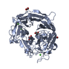
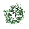
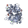
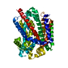
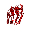
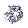
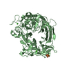
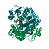
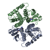
| Title | Crystal structure of polysaccharide lyase Uly1 | ||||||
 Components Components | Uly1 | ||||||
 Keywords Keywords | LYASE / ulvan lyase / polysaccharide lyase family 24 / marine bacterium | ||||||
| Function / homology | BNR repeat-containing family member / metal ion binding / Uncharacterized protein Function and homology information Function and homology information | ||||||
| Biological species |  Catenovulum maritimum (bacteria) Catenovulum maritimum (bacteria) | ||||||
| Method |  X-RAY DIFFRACTION / SYNCHROTRON / SAD / Resolution: 1.79 Å X-RAY DIFFRACTION / SYNCHROTRON / SAD / Resolution: 1.79 Å | ||||||
 Authors Authors | Chen, X.L. / Cao, H.Y. / Xu, F. / Dong, F. | ||||||
 Citation Citation | Journal: Appl.Environ.Microbiol. / Year: 2021 Title: Mechanistic Insights into Substrate Recognition and Catalysis of a New Ulvan Lyase of Polysaccharide Lyase Family 24. Authors: Xu, F. / Dong, F. / Sun, X.H. / Cao, H.Y. / Fu, H.H. / Li, C.Y. / Zhang, X.Y. / McMinn, A. / Zhang, Y.Z. / Wang, P. / Chen, X.L. | ||||||
| History |
|
- Structure visualization
| Structure viewer | Molecule: MolmilJmol/JSmol |
|---|
- Downloads & links
-Download
| PDBx/mmCIF format | 7drq.cif.gz | 237.8 KB | Display | PDBx/mmCIF format |
|---|---|---|---|---|
| PDB format | pdb7drq.ent.gz | 184.8 KB | Display | PDB format |
| PDBx/mmJSON format | 7drq.json.gz | Tree view | PDBx/mmJSON format | |
| Others |  Other downloads Other downloads |
-Validation report
| Arichive directory | https://data.pdbj.org/pub/pdb/validation_reports/dr/7drqftp://data.pdbj.org/pub/pdb/validation_reports/dr/7drq | HTTPS FTP |
|---|
-Related structure data


-Links
 PDBj
PDBj- Assembly
| Deposited unit | 
| ||||||||
|---|---|---|---|---|---|---|---|---|---|
| 1 | 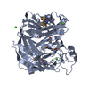
| ||||||||
| 2 | 
| ||||||||
| Unit cell |
|
-Components
| #1: Protein | Mass: 57404.820 Da / Num. of mol.: 2 Source method: isolated from a genetically manipulated source Source: (gene. exp.) Catenovulum maritimum (bacteria) / Gene: XM47_11230 / Production host: #2: Chemical | ChemComp-CA /   Mass: 40.078 Da / Num. of mol.: 5 / Source method: obtained synthetically / Formula: Ca Mass: 40.078 Da / Num. of mol.: 5 / Source method: obtained synthetically / Formula: Ca#3: Water | ChemComp-HOH / |  Mass: 18.015 Da / Num. of mol.: 1399 / Source method: isolated from a natural source / Formula: H2O Mass: 18.015 Da / Num. of mol.: 1399 / Source method: isolated from a natural source / Formula: H2OHas ligand of interest | N | Has protein modification | Y | |
|---|
-Experimental details
-Experiment
| Experiment | Method: X-RAY DIFFRACTION / Number of used crystals: 1 |
|---|
- Sample preparation
| Crystal | Density Matthews: 2.52 Å3/Da / Density % sol: 51.14 % Description: THE ENTRY CONTAINS FRIEDEL PAIRS IN I/F_PLUS/MINUS COLUMNS. |
|---|---|
| Crystal grow | Temperature: 293 K / Method: vapor diffusion, hanging drop Details: 200 mM calcium acetate, 100 mM MES (pH 6.0), 20% (w/v) PEG 8000 |
-Data collection
| Diffraction | Mean temperature: 100 K / Serial crystal experiment: N |
|---|---|
| Diffraction source | Source: SYNCHROTRON / Site: SSRF  / Beamline: BL17U / Wavelength: 0.9791 Å / Beamline: BL17U / Wavelength: 0.9791 Å |
| Detector | Type: ADSC QUANTUM 315r / Detector: CCD / Date: Jun 27, 2017 |
| Radiation | Protocol: SINGLE WAVELENGTH / Monochromatic (M) / Laue (L): M / Scattering type: x-ray |
| Radiation wavelength | Wavelength: 0.9791 Å / Relative weight: 1 |
| Reflection | Resolution: 1.79→37.11 Å / Num. obs: 101227 / % possible obs: 99.16 % / Redundancy: 6.8 % / Rmerge(I) obs: 0.118 / Rpim(I) all: 0.049 / Rrim(I) all: 0.128 / Net I/σ(I): 22.79 |
| Reflection shell | Resolution: 1.79→1.86 Å / Redundancy: 7 % / Rmerge(I) obs: 0.286 / Num. unique obs: 9595 / Rpim(I) all: 0.116 / Rrim(I) all: 0.309 / % possible all: 94.48 |
- Processing
| Software |
| |||||||||||||||||||||||||||||||||||||||||||||||||||||||||||||||||||||||||||||||||||||||||||||||||||||||||||||||||||||||||||||||||||||||||||||||||||||||||||||||||||||||||||||||||||||||||||||||||||||||||||||||||||||||||
|---|---|---|---|---|---|---|---|---|---|---|---|---|---|---|---|---|---|---|---|---|---|---|---|---|---|---|---|---|---|---|---|---|---|---|---|---|---|---|---|---|---|---|---|---|---|---|---|---|---|---|---|---|---|---|---|---|---|---|---|---|---|---|---|---|---|---|---|---|---|---|---|---|---|---|---|---|---|---|---|---|---|---|---|---|---|---|---|---|---|---|---|---|---|---|---|---|---|---|---|---|---|---|---|---|---|---|---|---|---|---|---|---|---|---|---|---|---|---|---|---|---|---|---|---|---|---|---|---|---|---|---|---|---|---|---|---|---|---|---|---|---|---|---|---|---|---|---|---|---|---|---|---|---|---|---|---|---|---|---|---|---|---|---|---|---|---|---|---|---|---|---|---|---|---|---|---|---|---|---|---|---|---|---|---|---|---|---|---|---|---|---|---|---|---|---|---|---|---|---|---|---|---|---|---|---|---|---|---|---|---|---|---|---|---|---|---|---|---|
| Refinement | Method to determine structure: SAD / Resolution: 1.79→37.11 Å / SU ML: 0.16 / Cross valid method: THROUGHOUT / σ(F): 1.38 / Phase error: 18.4 / Stereochemistry target values: ML
| |||||||||||||||||||||||||||||||||||||||||||||||||||||||||||||||||||||||||||||||||||||||||||||||||||||||||||||||||||||||||||||||||||||||||||||||||||||||||||||||||||||||||||||||||||||||||||||||||||||||||||||||||||||||||
| Solvent computation | Shrinkage radii: 0.9 Å / VDW probe radii: 1.11 Å / Solvent model: FLAT BULK SOLVENT MODEL | |||||||||||||||||||||||||||||||||||||||||||||||||||||||||||||||||||||||||||||||||||||||||||||||||||||||||||||||||||||||||||||||||||||||||||||||||||||||||||||||||||||||||||||||||||||||||||||||||||||||||||||||||||||||||
| Displacement parameters | Biso max: 54.05 Å2 / Biso mean: 19.1079 Å2 / Biso min: 4.89 Å2 | |||||||||||||||||||||||||||||||||||||||||||||||||||||||||||||||||||||||||||||||||||||||||||||||||||||||||||||||||||||||||||||||||||||||||||||||||||||||||||||||||||||||||||||||||||||||||||||||||||||||||||||||||||||||||
| Refinement step | Cycle: final / Resolution: 1.79→37.11 Å
| |||||||||||||||||||||||||||||||||||||||||||||||||||||||||||||||||||||||||||||||||||||||||||||||||||||||||||||||||||||||||||||||||||||||||||||||||||||||||||||||||||||||||||||||||||||||||||||||||||||||||||||||||||||||||
| LS refinement shell | Refine-ID: X-RAY DIFFRACTION / Rfactor Rfree error: 0 / Total num. of bins used: 30
|
