 Movie
Movie Controller

[English] 日本語
 Yorodumi
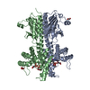
- PDB-7dog: Crystal structure of a nuclease and capping domain of SbcD from S... -
+ Open data

- Basic information
| Entry | Database: PDB / ID: 7dog | ||||||
|---|---|---|---|---|---|---|---|
| Title | Crystal structure of a nuclease and capping domain of SbcD from Staphylococcus aureus | ||||||
 Components Components | Nuclease SbcCD subunit D | ||||||
 Keywords Keywords | HYDROLASE / DNA nuclease / endonuclease / exonuclease / DNA repair | ||||||
| Function / homology |  Function and homology information Function and homology information3'-5' exonuclease activity / endonuclease activity / DNA recombination / DNA replication Similarity search - Function | ||||||
| Biological species |  Staphylococcus aureus subsp. aureus Mu50 (bacteria) Staphylococcus aureus subsp. aureus Mu50 (bacteria) | ||||||
| Method |  X-RAY DIFFRACTION / SYNCHROTRON / MOLECULAR REPLACEMENT / Resolution: 2.91 Å X-RAY DIFFRACTION / SYNCHROTRON / MOLECULAR REPLACEMENT / Resolution: 2.91 Å | ||||||
 Authors Authors | Lee, J. / Ha, N.-C. | ||||||
 Citation Citation | Journal: J.Microbiol / Year: 2021 Title: Crystal structure of the nuclease and capping domain of SbcD from Staphylococcus aureus. Authors: Lee, J. / Jo, I. / Ahn, J. / Hong, S. / Jeong, S. / Kwon, A. / Ha, N.C. | ||||||
| History |
|
- Structure visualization
| Structure viewer | Molecule: MolmilJmol/JSmol |
|---|
- Downloads & links
-Download
| PDBx/mmCIF format | 7dog.cif.gz | 168.7 KB | Display | PDBx/mmCIF format |
|---|---|---|---|---|
| PDB format | pdb7dog.ent.gz | 107.3 KB | Display | PDB format |
| PDBx/mmJSON format | 7dog.json.gz | Tree view | PDBx/mmJSON format | |
| Others |  Other downloads Other downloads |
-Validation report
| Arichive directory | https://data.pdbj.org/pub/pdb/validation_reports/do/7dogftp://data.pdbj.org/pub/pdb/validation_reports/do/7dog | HTTPS FTP |
|---|
-Related structure data
| Related structure data |  6s6vS S: Starting model for refinement |
|---|---|
| Similar structure data |


-Links
 PDBj
PDBj
- Assembly
| Deposited unit | 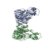
| ||||||||||||
|---|---|---|---|---|---|---|---|---|---|---|---|---|---|
| 1 |
| ||||||||||||
| Unit cell |
|
-Components
| #1: Protein | Mass: 36819.805 Da / Num. of mol.: 2 Source method: isolated from a genetically manipulated source Source: (gene. exp.) Staphylococcus aureus subsp. aureus Mu50 (bacteria)Strain: Mu50 / Gene: sbcD, SAV1345 / Production host: #2: Chemical | ChemComp-MN /   Mass: 54.938 Da / Num. of mol.: 4 / Source method: obtained synthetically / Formula: Mn / Feature type: SUBJECT OF INVESTIGATION Mass: 54.938 Da / Num. of mol.: 4 / Source method: obtained synthetically / Formula: Mn / Feature type: SUBJECT OF INVESTIGATIONHas ligand of interest | Y | |
|---|
-Experimental details
-Experiment
| Experiment | Method: X-RAY DIFFRACTION / Number of used crystals: 1 |
|---|
- Sample preparation
| Crystal | Density Matthews: 2.62 Å3/Da / Density % sol: 53 % |
|---|---|
| Crystal grow | Temperature: 287.15 K / Method: vapor diffusion, hanging drop Details: 2% (v/v) TacsimateTM (pH 6.0), 0.1 M Bis-Tris (pH 7.0), 18% (w/v) polyethylene glycol 3350 |
-Data collection
| Diffraction | Mean temperature: 80 K / Serial crystal experiment: N |
|---|---|
| Diffraction source | Source: SYNCHROTRON / Site: PAL/PLS  / Beamline: 11C / Wavelength: 0.9794 Å / Beamline: 11C / Wavelength: 0.9794 Å |
| Detector | Type: DECTRIS PILATUS3 6M / Detector: PIXEL / Date: Dec 18, 2019 |
| Radiation | Protocol: SINGLE WAVELENGTH / Monochromatic (M) / Laue (L): M / Scattering type: x-ray |
| Radiation wavelength | Wavelength: 0.9794 Å / Relative weight: 1 |
| Reflection | Resolution: 2.9→50 Å / Num. obs: 16408 / % possible obs: 93.8 % / Redundancy: 5.5 % / Biso Wilson estimate: 46.69 Å2 / CC1/2: 0.989 / CC star: 0.997 / Rmerge(I) obs: 0.148 / Rpim(I) all: 0.051 / Rrim(I) all: 0.143 / Net I/σ(I): 10.7 |
| Reflection shell | Resolution: 2.9→2.95 Å / Redundancy: 3.5 % / Rmerge(I) obs: 0.333 / Mean I/σ(I) obs: 3.5 / Num. unique obs: 749 / CC1/2: 0.752 / CC star: 0.926 / Rpim(I) all: 0.138 / Rrim(I) all: 0.297 / % possible all: 86.7 |
- Processing
| Software |
| |||||||||||||||||||||||||||||||||||||||||||||||||
|---|---|---|---|---|---|---|---|---|---|---|---|---|---|---|---|---|---|---|---|---|---|---|---|---|---|---|---|---|---|---|---|---|---|---|---|---|---|---|---|---|---|---|---|---|---|---|---|---|---|---|
| Refinement | Method to determine structure: MOLECULAR REPLACEMENT Starting model: 6s6v Resolution: 2.91→48.84 Å / SU ML: 0.2667 / Cross valid method: FREE R-VALUE / σ(F): 1.59 / Phase error: 21.0164 Stereochemistry target values: GeoStd + Monomer Library + CDL v1.2
| |||||||||||||||||||||||||||||||||||||||||||||||||
| Solvent computation | Shrinkage radii: 0.9 Å / VDW probe radii: 1.11 Å / Solvent model: FLAT BULK SOLVENT MODEL | |||||||||||||||||||||||||||||||||||||||||||||||||
| Displacement parameters | Biso mean: 43.64 Å2 | |||||||||||||||||||||||||||||||||||||||||||||||||
| Refinement step | Cycle: LAST / Resolution: 2.91→48.84 Å
| |||||||||||||||||||||||||||||||||||||||||||||||||
| Refine LS restraints |
| |||||||||||||||||||||||||||||||||||||||||||||||||
| LS refinement shell |
|
