 Movie
Movie Controller

[English] 日本語
 Yorodumi
- PDB-7djm: Structure of four truncated and mutated forms of quenching protein -
+ Open data

- Basic information
| Entry | Database: PDB / ID: 7djm | |||||||||||||||||||||
|---|---|---|---|---|---|---|---|---|---|---|---|---|---|---|---|---|---|---|---|---|---|---|
| Title | Structure of four truncated and mutated forms of quenching protein | |||||||||||||||||||||
 Components Components | Protein SUPPRESSOR OF QUENCHING 1, chloroplastic | |||||||||||||||||||||
 Keywords Keywords | PLANT PROTEIN / Suppressor / Quenching | |||||||||||||||||||||
| Function / homology |  Function and homology information Function and homology informationnonphotochemical quenching / thylakoid membrane / chloroplast thylakoid / Hydrolases; Acting on ester bonds; Phosphoric-monoester hydrolases / chloroplast stroma / chloroplast thylakoid membrane / chloroplast / hydrolase activity / metal ion binding / nucleus Similarity search - Function | |||||||||||||||||||||
| Biological species |  | |||||||||||||||||||||
| Method |  X-RAY DIFFRACTION / SYNCHROTRON / MOLECULAR REPLACEMENT / Resolution: 1.70000117792 Å X-RAY DIFFRACTION / SYNCHROTRON / MOLECULAR REPLACEMENT / Resolution: 1.70000117792 Å | |||||||||||||||||||||
 Authors Authors | Yu, G.M. / Pan, X.W. / Li, M. | |||||||||||||||||||||
| Funding support |  China, 6items China, 6items
| |||||||||||||||||||||
 Citation Citation | Journal: Nat.Plants / Year: 2022 Title: Structure of Arabidopsis SOQ1 lumenal region unveils C-terminal domain essential for negative regulation of photoprotective qH. Authors: Yu, G. / Hao, J. / Pan, X. / Shi, L. / Zhang, Y. / Wang, J. / Fan, H. / Xiao, Y. / Yang, F. / Lou, J. / Chang, W. / Malnoe, A. / Li, M. | |||||||||||||||||||||
| History |
|
- Structure visualization
| Structure viewer | Molecule: MolmilJmol/JSmol |
|---|
- Downloads & links
-Download
| PDBx/mmCIF format | 7djm.cif.gz | 116.4 KB | Display | PDBx/mmCIF format |
|---|---|---|---|---|
| PDB format | pdb7djm.ent.gz | 82.2 KB | Display | PDB format |
| PDBx/mmJSON format | 7djm.json.gz | Tree view | PDBx/mmJSON format | |
| Others |  Other downloads Other downloads |
-Validation report
| Arichive directory | https://data.pdbj.org/pub/pdb/validation_reports/dj/7djmftp://data.pdbj.org/pub/pdb/validation_reports/dj/7djm | HTTPS FTP |
|---|
-Related structure data
| Related structure data | 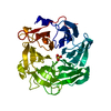 7djjSC 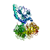 7djkC  7djlC S: Starting model for refinement C: citing same article ( |
|---|---|
| Similar structure data |
-Links
 PDBj
PDBj


- Assembly
| Deposited unit | 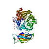
| ||||||||||||
|---|---|---|---|---|---|---|---|---|---|---|---|---|---|
| 1 |
| ||||||||||||
| Unit cell |
|
-Components
| #1: Protein | Mass: 54123.477 Da / Num. of mol.: 1 / Fragment: NHL_CTD Source method: isolated from a genetically manipulated source Source: (gene. exp.)  Komagataella phaffii GS115 (fungus) Komagataella phaffii GS115 (fungus)References: UniProt: Q8VZ10, Hydrolases; Acting on ester bonds; Phosphoric-monoester hydrolases | ||||||||
|---|---|---|---|---|---|---|---|---|---|
| #2: Chemical |   Mass: 59.044 Da / Num. of mol.: 2 / Source method: isolated from a natural source / Formula: C2H3O2 / Feature type: SUBJECT OF INVESTIGATION Mass: 59.044 Da / Num. of mol.: 2 / Source method: isolated from a natural source / Formula: C2H3O2 / Feature type: SUBJECT OF INVESTIGATION#3: Chemical | ChemComp-DTT / |   Mass: 154.251 Da / Num. of mol.: 1 / Source method: obtained synthetically / Formula: C4H10O2S2 / Feature type: SUBJECT OF INVESTIGATION Mass: 154.251 Da / Num. of mol.: 1 / Source method: obtained synthetically / Formula: C4H10O2S2 / Feature type: SUBJECT OF INVESTIGATION#4: Chemical | ChemComp-NA / |   Mass: 22.990 Da / Num. of mol.: 1 / Source method: obtained synthetically / Formula: Na / Feature type: SUBJECT OF INVESTIGATION Mass: 22.990 Da / Num. of mol.: 1 / Source method: obtained synthetically / Formula: Na / Feature type: SUBJECT OF INVESTIGATION#5: Water | ChemComp-HOH / |  Mass: 18.015 Da / Num. of mol.: 284 / Source method: isolated from a natural source / Formula: H2O Mass: 18.015 Da / Num. of mol.: 284 / Source method: isolated from a natural source / Formula: H2OHas ligand of interest | Y | |
-Experimental details
-Experiment
| Experiment | Method: X-RAY DIFFRACTION / Number of used crystals: 1 |
|---|
- Sample preparation
| Crystal | Density Matthews: 2.06 Å3/Da / Density % sol: 40.32 % |
|---|---|
| Crystal grow | Temperature: 291.15 K / Method: vapor diffusion, sitting drop / Details: 25-34% PEG 3350, 0.2 M NH4Ac, 0.1M MES, pH 5.9-6.6 / PH range: 5.9-6.6 |
-Data collection
| Diffraction | Mean temperature: 80 K / Serial crystal experiment: N |
|---|---|
| Diffraction source | Source: SYNCHROTRON / Site: SSRF / Beamline: BL19U1 / Wavelength: 0.9777 Å |
| Detector | Type: DECTRIS PILATUS3 X 6M / Detector: PIXEL / Date: Dec 16, 2017 |
| Radiation | Protocol: SINGLE WAVELENGTH / Monochromatic (M) / Laue (L): M / Scattering type: x-ray |
| Radiation wavelength | Wavelength: 0.9777 Å / Relative weight: 1 |
| Reflection | Resolution: 1.7→50 Å / Num. obs: 49848 / % possible obs: 99.8 % / Redundancy: 12.4 % / Biso Wilson estimate: 22.0080859116 Å2 / Rrim(I) all: 0.082 / Net I/σ(I): 33.36 |
| Reflection shell | Resolution: 1.7→1.73 Å / Num. unique obs: 2367 / CC1/2: 0.864 |
- Processing
| Software |
| |||||||||||||||||||||||||||||||||||||||||||||||||||||||||||||||||||||||||||||||||||||||||||||||||||||||||||||||||||||||||||||||||||||
|---|---|---|---|---|---|---|---|---|---|---|---|---|---|---|---|---|---|---|---|---|---|---|---|---|---|---|---|---|---|---|---|---|---|---|---|---|---|---|---|---|---|---|---|---|---|---|---|---|---|---|---|---|---|---|---|---|---|---|---|---|---|---|---|---|---|---|---|---|---|---|---|---|---|---|---|---|---|---|---|---|---|---|---|---|---|---|---|---|---|---|---|---|---|---|---|---|---|---|---|---|---|---|---|---|---|---|---|---|---|---|---|---|---|---|---|---|---|---|---|---|---|---|---|---|---|---|---|---|---|---|---|---|---|---|
| Refinement | Method to determine structure: MOLECULAR REPLACEMENT Starting model: 7DJJ Resolution: 1.70000117792→39.375 Å / SU ML: 0.167139620693 / Cross valid method: FREE R-VALUE / σ(F): 1.36333929852 / Phase error: 19.4162754482 Stereochemistry target values: GeoStd + Monomer Library + CDL v1.2
| |||||||||||||||||||||||||||||||||||||||||||||||||||||||||||||||||||||||||||||||||||||||||||||||||||||||||||||||||||||||||||||||||||||
| Solvent computation | Shrinkage radii: 0.9 Å / VDW probe radii: 1.11 Å / Solvent model: FLAT BULK SOLVENT MODEL | |||||||||||||||||||||||||||||||||||||||||||||||||||||||||||||||||||||||||||||||||||||||||||||||||||||||||||||||||||||||||||||||||||||
| Displacement parameters | Biso mean: 31.9819224725 Å2 | |||||||||||||||||||||||||||||||||||||||||||||||||||||||||||||||||||||||||||||||||||||||||||||||||||||||||||||||||||||||||||||||||||||
| Refinement step | Cycle: LAST / Resolution: 1.70000117792→39.375 Å
| |||||||||||||||||||||||||||||||||||||||||||||||||||||||||||||||||||||||||||||||||||||||||||||||||||||||||||||||||||||||||||||||||||||
| Refine LS restraints |
| |||||||||||||||||||||||||||||||||||||||||||||||||||||||||||||||||||||||||||||||||||||||||||||||||||||||||||||||||||||||||||||||||||||
| LS refinement shell |
|
