 Movie
Movie Controller

[English] 日本語
 Yorodumi
- PDB-7dib: Crystal structure of D-threonine aldolase from Filomicrobium marinum -
+ Open data

- Basic information
| Entry | Database: PDB / ID: 7dib | ||||||
|---|---|---|---|---|---|---|---|
| Title | Crystal structure of D-threonine aldolase from Filomicrobium marinum | ||||||
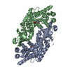
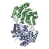
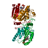
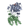
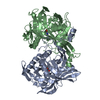
 Components Components | D-threonine aldolase | ||||||
 Keywords Keywords | BIOSYNTHETIC PROTEIN / aldolase / PLP-dependent enzyme | ||||||
| Function / homology |  Function and homology information Function and homology informationD-threonine aldolase / D-threonine aldolase activity / D-serine ammonia-lyase activity / D-serine catabolic process Similarity search - Function | ||||||
| Biological species |  Candidatus Filomicrobium marinum (bacteria) Candidatus Filomicrobium marinum (bacteria) | ||||||
| Method |  X-RAY DIFFRACTION / SYNCHROTRON / MOLECULAR REPLACEMENT / molecular replacement / Resolution: 2.2 Å X-RAY DIFFRACTION / SYNCHROTRON / MOLECULAR REPLACEMENT / molecular replacement / Resolution: 2.2 Å | ||||||
 Authors Authors | Seo, H. / Kim, K.-J. | ||||||
 Citation Citation | Journal: Acs Catalysis / Year: 2021 Title: Cbeta-Selective Aldol Addition of d-Threonine Aldolase by Spatial Constraint of Aldehyde Binding. Authors: Park, S.-H. / Seo, H. / Seok, J. / Kim, H. / Kwon, K.K. / Yeom, S.-J. / Lee, S.-G. / Kim, K.-J. | ||||||
| History |
|
- Structure visualization
| Structure viewer | Molecule: MolmilJmol/JSmol |
|---|
- Downloads & links
-Download
| PDBx/mmCIF format | 7dib.cif.gz | 150.7 KB | Display | PDBx/mmCIF format |
|---|---|---|---|---|
| PDB format | pdb7dib.ent.gz | 116.7 KB | Display | PDB format |
| PDBx/mmJSON format | 7dib.json.gz | Tree view | PDBx/mmJSON format | |
| Others |  Other downloads Other downloads |
-Validation report
| Summary document | 7dib_validation.pdf.gz | 453.6 KB | Display | wwPDB validaton report |
|---|---|---|---|---|
| Full document | 7dib_full_validation.pdf.gz | 460.8 KB | Display | |
| Data in XML | 7dib_validation.xml.gz | 27.7 KB | Display | |
| Data in CIF | 7dib_validation.cif.gz | 37.9 KB | Display | |
| Arichive directory | https://data.pdbj.org/pub/pdb/validation_reports/di/7dibftp://data.pdbj.org/pub/pdb/validation_reports/di/7dib | HTTPS FTP |
-Related structure data
| Related structure data | 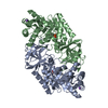 4v15S S: Starting model for refinement |
|---|---|
| Similar structure data |


-Links
 PDBj
PDBj- Assembly
| Deposited unit | 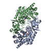
| ||||||||
|---|---|---|---|---|---|---|---|---|---|
| 1 |
| ||||||||
| Unit cell |
|
-Components
| #1: Protein | Mass: 42495.023 Da / Num. of mol.: 2 Source method: isolated from a genetically manipulated source Details: The sequence "MGSSHHHHHHSSGLVPRGSH" are derived from pET28a vector. Source: (gene. exp.) Candidatus Filomicrobium marinum (bacteria)Gene: YBN1229_v1_3680 / Plasmid: pET28a / Production host: #2: Water | ChemComp-HOH / |  Mass: 18.015 Da / Num. of mol.: 64 / Source method: isolated from a natural source / Formula: H2O Mass: 18.015 Da / Num. of mol.: 64 / Source method: isolated from a natural source / Formula: H2OHas ligand of interest | Y | |
|---|
-Experimental details
-Experiment
| Experiment | Method: X-RAY DIFFRACTION / Number of used crystals: 1 |
|---|
- Sample preparation
| Crystal | Density Matthews: 2.43 Å3/Da / Density % sol: 49.44 % |
|---|---|
| Crystal grow | Temperature: 298 K / Method: vapor diffusion, sitting drop / pH: 4.6 Details: PEG 400, Sodium acetate tribasic, Cadmium chloride hydrate |
-Data collection
| Diffraction | Mean temperature: 100 K / Serial crystal experiment: N |
|---|---|
| Diffraction source | Source: SYNCHROTRON / Site: PAL/PLS  / Beamline: 7A (6B, 6C1) / Wavelength: 0.97934 Å / Beamline: 7A (6B, 6C1) / Wavelength: 0.97934 Å |
| Detector | Type: ADSC QUANTUM 270 / Detector: CCD / Date: Sep 17, 2018 |
| Radiation | Monochromator: Double Crystal Monochromator / Protocol: SINGLE WAVELENGTH / Monochromatic (M) / Laue (L): M / Scattering type: x-ray |
| Radiation wavelength | Wavelength: 0.97934 Å / Relative weight: 1 |
| Reflection | Resolution: 2.2→50 Å / Num. obs: 41469 / % possible obs: 96.1 % / Redundancy: 4 % / CC1/2: 0.982 / Rmerge(I) obs: 0.082 / Net I/σ(I): 19.3 |
| Reflection shell | Resolution: 2.2→2.24 Å / Rmerge(I) obs: 0.398 / Mean I/σ(I) obs: 3.3 / Num. unique obs: 1981 / CC1/2: 0.585 |
-Phasing
| Phasing | Method: molecular replacement |
|---|
- Processing
| Software |
| ||||||||||||||||||||||||||||||||||||||||||||||||||||||||||||
|---|---|---|---|---|---|---|---|---|---|---|---|---|---|---|---|---|---|---|---|---|---|---|---|---|---|---|---|---|---|---|---|---|---|---|---|---|---|---|---|---|---|---|---|---|---|---|---|---|---|---|---|---|---|---|---|---|---|---|---|---|---|
| Refinement | Method to determine structure: MOLECULAR REPLACEMENT Starting model: 4v15 Resolution: 2.2→31.3 Å / Cor.coef. Fo:Fc: 0.935 / Cor.coef. Fo:Fc free: 0.907 / SU B: 7.593 / SU ML: 0.183 / Cross valid method: THROUGHOUT / σ(F): 0 / ESU R: 0.276 / ESU R Free: 0.217 / Stereochemistry target values: MAXIMUM LIKELIHOOD Details: HYDROGENS HAVE BEEN ADDED IN THE RIDING POSITIONS U VALUES : REFINED INDIVIDUALLY
| ||||||||||||||||||||||||||||||||||||||||||||||||||||||||||||
| Solvent computation | Ion probe radii: 0.8 Å / Shrinkage radii: 0.8 Å / VDW probe radii: 1.2 Å / Solvent model: MASK | ||||||||||||||||||||||||||||||||||||||||||||||||||||||||||||
| Displacement parameters | Biso max: 125.36 Å2 / Biso mean: 31.372 Å2 / Biso min: 12.8 Å2
| ||||||||||||||||||||||||||||||||||||||||||||||||||||||||||||
| Refinement step | Cycle: final / Resolution: 2.2→31.3 Å
| ||||||||||||||||||||||||||||||||||||||||||||||||||||||||||||
| Refine LS restraints |
| ||||||||||||||||||||||||||||||||||||||||||||||||||||||||||||
| LS refinement shell | Resolution: 2.2→2.253 Å / Rfactor Rfree error: 0
|
