 Movie
Movie Controller


+ Open data

- Basic information
| Entry | Database: PDB / ID: 7dhd | ||||||
|---|---|---|---|---|---|---|---|
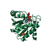
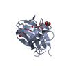
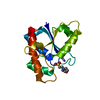
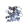
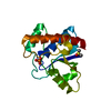
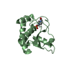
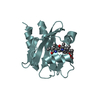
| Title | Vibrio vulnificus Wzb | ||||||
 Components Components | Protein-tyrosine-phosphatase | ||||||
 Keywords Keywords | HYDROLASE / Vibrio / Wzb / low molecular weight protein tyrosine phosphatase (LMWPTP) / capsular polysaccharide | ||||||
| Function / homology | : / Protein-tyrosine phosphatase, low molecular weight / Phosphotyrosine protein phosphatase I / Phosphotyrosine protein phosphatase I superfamily / Low molecular weight phosphotyrosine protein phosphatase / Low molecular weight phosphatase family / protein-tyrosine-phosphatase / protein tyrosine phosphatase activity / protein-tyrosine-phosphatase Function and homology information Function and homology information | ||||||
| Biological species |  Vibrio vulnificus (bacteria) Vibrio vulnificus (bacteria) | ||||||
| Method |  X-RAY DIFFRACTION / SYNCHROTRON / MOLECULAR REPLACEMENT / Resolution: 1.71 Å X-RAY DIFFRACTION / SYNCHROTRON / MOLECULAR REPLACEMENT / Resolution: 1.71 Å | ||||||
 Authors Authors | Ma, Q. / Wang, X. | ||||||
| Funding support |  China, 1items China, 1items
| ||||||
 Citation Citation | Journal: J.Biol.Chem. / Year: 2021 Title: Wzb of Vibrio vulnificus represents a new group of low-molecular-weight protein tyrosine phosphatases with a unique insertion in the W-loop. Authors: Wang, X. / Ma, Q. | ||||||
| History |
|
- Structure visualization
| Structure viewer | Molecule: MolmilJmol/JSmol |
|---|
- Downloads & links
-Download
| PDBx/mmCIF format | 7dhd.cif.gz | 133.6 KB | Display | PDBx/mmCIF format |
|---|---|---|---|---|
| PDB format | pdb7dhd.ent.gz | 101.7 KB | Display | PDB format |
| PDBx/mmJSON format | 7dhd.json.gz | Tree view | PDBx/mmJSON format | |
| Others |  Other downloads Other downloads |
-Validation report
| Arichive directory | https://data.pdbj.org/pub/pdb/validation_reports/dh/7dhdftp://data.pdbj.org/pub/pdb/validation_reports/dh/7dhd | HTTPS FTP |
|---|
-Related structure data
| Related structure data |  7dheC  7dhfC  2wmyS S: Starting model for refinement C: citing same article ( |
|---|---|
| Similar structure data |


-Links
 PDBj
PDBj
- Assembly
| Deposited unit | 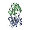
| ||||||||
|---|---|---|---|---|---|---|---|---|---|
| 1 | 
| ||||||||
| 2 | 
| ||||||||
| Unit cell |
|
-Components
| #1: Protein | Mass: 16314.879 Da / Num. of mol.: 2 Source method: isolated from a genetically manipulated source Source: (gene. exp.) Vibrio vulnificus (bacteria) / Gene: wzb / Production host: #2: Chemical |   Mass: 35.453 Da / Num. of mol.: 2 / Source method: obtained synthetically / Formula: Cl Mass: 35.453 Da / Num. of mol.: 2 / Source method: obtained synthetically / Formula: Cl#3: Water | ChemComp-HOH / |  Mass: 18.015 Da / Num. of mol.: 270 / Source method: isolated from a natural source / Formula: H2O Mass: 18.015 Da / Num. of mol.: 270 / Source method: isolated from a natural source / Formula: H2OHas ligand of interest | N | |
|---|
-Experimental details
-Experiment
| Experiment | Method: X-RAY DIFFRACTION / Number of used crystals: 1 |
|---|
- Sample preparation
| Crystal | Density Matthews: 2.07 Å3/Da / Density % sol: 40.66 % |
|---|---|
| Crystal grow | Temperature: 293 K / Method: vapor diffusion, sitting drop Details: Free VvWzb crystals were grown in drops containing 1.5 ul of protein solution (14 mg/ml in the buffer 10 mM HEPES pH 7.5, 150 mM NaCl, 1 mM DTT) and 1.5 ul of reservoir solution (100 mM ...Details: Free VvWzb crystals were grown in drops containing 1.5 ul of protein solution (14 mg/ml in the buffer 10 mM HEPES pH 7.5, 150 mM NaCl, 1 mM DTT) and 1.5 ul of reservoir solution (100 mM HEPES pH7.5, 200 mM NaCl, 31% (w/v) PEG 3350) |
-Data collection
| Diffraction | Mean temperature: 100 K / Serial crystal experiment: N |
|---|---|
| Diffraction source | Source: SYNCHROTRON / Site: SSRF / Beamline: BL19U1 / Wavelength: 0.9788 Å |
| Detector | Type: DECTRIS PILATUS3 6M / Detector: PIXEL / Date: Nov 11, 2019 |
| Radiation | Protocol: SINGLE WAVELENGTH / Monochromatic (M) / Laue (L): M / Scattering type: x-ray |
| Radiation wavelength | Wavelength: 0.9788 Å / Relative weight: 1 |
| Reflection | Resolution: 1.71→58.66 Å / Num. obs: 29957 / % possible obs: 99.5 % / Redundancy: 12.6 % / CC1/2: 0.999 / Rpim(I) all: 0.025 / Rrim(I) all: 0.088 / Rsym value: 0.085 / Net I/σ(I): 19.2 |
| Reflection shell | Resolution: 1.711→1.74 Å / Redundancy: 12.9 % / Mean I/σ(I) obs: 2.3 / Num. unique obs: 1506 / CC1/2: 0.788 / Rpim(I) all: 0.343 / Rrim(I) all: 1.238 / Rsym value: 1.188 / % possible all: 99.7 |
- Processing
| Software |
| ||||||||||||||||||||||||||||||||||||||||||||||||||||||||||||||||||||||||||||||||||||||||||||||||||||||||||||
|---|---|---|---|---|---|---|---|---|---|---|---|---|---|---|---|---|---|---|---|---|---|---|---|---|---|---|---|---|---|---|---|---|---|---|---|---|---|---|---|---|---|---|---|---|---|---|---|---|---|---|---|---|---|---|---|---|---|---|---|---|---|---|---|---|---|---|---|---|---|---|---|---|---|---|---|---|---|---|---|---|---|---|---|---|---|---|---|---|---|---|---|---|---|---|---|---|---|---|---|---|---|---|---|---|---|---|---|---|---|
| Refinement | Method to determine structure: MOLECULAR REPLACEMENT Starting model: 2WMY Resolution: 1.71→58.66 Å / Cor.coef. Fo:Fc: 0.925 / Cor.coef. Fo:Fc free: 0.92 / SU R Cruickshank DPI: 0.122 / Cross valid method: THROUGHOUT / σ(F): 0 / SU R Blow DPI: 0.133 / SU Rfree Blow DPI: 0.117 / SU Rfree Cruickshank DPI: 0.111
| ||||||||||||||||||||||||||||||||||||||||||||||||||||||||||||||||||||||||||||||||||||||||||||||||||||||||||||
| Displacement parameters | Biso max: 111.09 Å2 / Biso mean: 32.58 Å2 / Biso min: 15.31 Å2
| ||||||||||||||||||||||||||||||||||||||||||||||||||||||||||||||||||||||||||||||||||||||||||||||||||||||||||||
| Refine analyze | Luzzati coordinate error obs: 0.26 Å | ||||||||||||||||||||||||||||||||||||||||||||||||||||||||||||||||||||||||||||||||||||||||||||||||||||||||||||
| Refinement step | Cycle: final / Resolution: 1.71→58.66 Å
| ||||||||||||||||||||||||||||||||||||||||||||||||||||||||||||||||||||||||||||||||||||||||||||||||||||||||||||
| Refine LS restraints |
| ||||||||||||||||||||||||||||||||||||||||||||||||||||||||||||||||||||||||||||||||||||||||||||||||||||||||||||
| LS refinement shell | Resolution: 1.71→1.77 Å / Rfactor Rfree error: 0 / Total num. of bins used: 15
| ||||||||||||||||||||||||||||||||||||||||||||||||||||||||||||||||||||||||||||||||||||||||||||||||||||||||||||
| Refinement TLS params. | Method: refined / Refine-ID: X-RAY DIFFRACTION
| ||||||||||||||||||||||||||||||||||||||||||||||||||||||||||||||||||||||||||||||||||||||||||||||||||||||||||||
| Refinement TLS group |
|
