 Movie
Movie Controller

[English] 日本語
 Yorodumi
- PDB-7dfs: Crystal structure of a novel 4-O-alpha-L-rhamnosyl-beta-D-glucuro... -
+ Open data

- Basic information
| Entry | Database: PDB / ID: 7dfs | ||||||
|---|---|---|---|---|---|---|---|
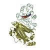
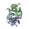
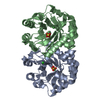
| Title | Crystal structure of a novel 4-O-alpha-L-rhamnosyl-beta-D-glucuronidase from Fusarium oxysporum 12S - Rha-GlcA complex | ||||||
 Components Components | 4-O-alpha-L-rhamnosyl-beta-D-glucuronidase | ||||||
 Keywords Keywords | HYDROLASE / TIM barrel / antiparallel beta-sheet | ||||||
| Function / homology | Golgi alpha-mannosidase II / Glycosidases / TIM Barrel / Alpha-Beta Barrel / Immunoglobulin-like / Sandwich / Mainly Beta / Alpha Beta / alpha-D-mannopyranose Function and homology information Function and homology information | ||||||
| Biological species |  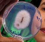 Fusarium oxysporum (fungus) Fusarium oxysporum (fungus) | ||||||
| Method |  X-RAY DIFFRACTION / SYNCHROTRON / MOLECULAR REPLACEMENT / Resolution: 1.49 Å X-RAY DIFFRACTION / SYNCHROTRON / MOLECULAR REPLACEMENT / Resolution: 1.49 Å | ||||||
 Authors Authors | Kondo, T. / Arakawa, T. / Fushinobu, S. / Sakamoto, T. | ||||||
 Citation Citation | Journal: Febs J. / Year: 2021 Title: Biochemical and structural characterization of a novel 4-O-alpha-l-rhamnosyl-beta-d-glucuronidase from Fusarium oxysporum. Authors: Kondo, T. / Kichijo, M. / Nakaya, M. / Takenaka, S. / Arakawa, T. / Kotake, T. / Fushinobu, S. / Sakamoto, T. | ||||||
| History |
|
- Structure visualization
| Structure viewer | Molecule: MolmilJmol/JSmol |
|---|
- Downloads & links
-Download
| PDBx/mmCIF format | 7dfs.cif.gz | 117.1 KB | Display | PDBx/mmCIF format |
|---|---|---|---|---|
| PDB format | pdb7dfs.ent.gz | 85.7 KB | Display | PDB format |
| PDBx/mmJSON format | 7dfs.json.gz | Tree view | PDBx/mmJSON format | |
| Others |  Other downloads Other downloads |
-Validation report
| Summary document | 7dfs_validation.pdf.gz | 1.7 MB | Display | wwPDB validaton report |
|---|---|---|---|---|
| Full document | 7dfs_full_validation.pdf.gz | 1.7 MB | Display | |
| Data in XML | 7dfs_validation.xml.gz | 23.8 KB | Display | |
| Data in CIF | 7dfs_validation.cif.gz | 37.2 KB | Display | |
| Arichive directory | https://data.pdbj.org/pub/pdb/validation_reports/df/7dfsftp://data.pdbj.org/pub/pdb/validation_reports/df/7dfs | HTTPS FTP |
-Related structure data


-Links
 PDBj
PDBj


- Assembly
| Deposited unit | 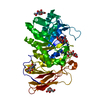
| ||||||||
|---|---|---|---|---|---|---|---|---|---|
| 1 |
| ||||||||
| Unit cell |
|
-Components
-Protein / Non-polymers , 2 types, 547 molecules A
| #1: Protein | Mass: 50763.793 Da / Num. of mol.: 1 Source method: isolated from a genetically manipulated source Source: (gene. exp.) Fusarium oxysporum (fungus) / Strain: 12S / Gene: FobglcA / Plasmid: pPICZ-alpha-A / Production host: Komagataella pastoris (fungus) / Strain (production host): X-33 / References: beta-glucuronidase |
|---|---|
| #6: Water | ChemComp-HOH / Mass: 18.015 Da / Num. of mol.: 546 / Source method: isolated from a natural source / Formula: H2O |
-Sugars , 4 types, 5 molecules 

| #2: Polysaccharide | alpha-L-rhamnopyranose-(1-4)-beta-D-glucopyranuronic acid Source method: isolated from a genetically manipulated source | ||
|---|---|---|---|
| #3: Polysaccharide | alpha-L-rhamnopyranose-(1-4)-alpha-D-glucopyranuronic acid Source method: isolated from a genetically manipulated source | ||
| #4: Sugar |  Type: D-saccharide, beta linking / Mass: 221.208 Da / Num. of mol.: 2 / Source method: obtained synthetically / Formula: C8H15NO6 / Feature type: SUBJECT OF INVESTIGATION Type: D-saccharide, beta linking / Mass: 221.208 Da / Num. of mol.: 2 / Source method: obtained synthetically / Formula: C8H15NO6 / Feature type: SUBJECT OF INVESTIGATION#5: Sugar | ChemComp-MAN / |  Type: D-saccharide, alpha linking / Mass: 180.156 Da / Num. of mol.: 1 / Source method: obtained synthetically / Formula: C6H12O6 / Feature type: SUBJECT OF INVESTIGATION Type: D-saccharide, alpha linking / Mass: 180.156 Da / Num. of mol.: 1 / Source method: obtained synthetically / Formula: C6H12O6 / Feature type: SUBJECT OF INVESTIGATION |
-Details
| Has ligand of interest | Y |
|---|---|
| Has protein modification | Y |
| Sequence details | The amino acid sequence of 4-O-alpha-L-rhamnosyl-beta-D-glucuronidase (FoBGlcA) has been registered ...The amino acid sequence of 4-O-alpha-L-rhamnosyl-beta-D-glucuronidase (FoBGlcA) has been registered in GenBank, DDBj and EMBL. Its accession number is "LC534636". |
-Experimental details
-Experiment
| Experiment | Method: X-RAY DIFFRACTION / Number of used crystals: 1 |
|---|
- Sample preparation
| Crystal | Density Matthews: 2.23 Å3/Da / Density % sol: 44.75 % |
|---|---|
| Crystal grow | Temperature: 293.15 K / Method: vapor diffusion, sitting drop / pH: 6 Details: 30% (v/v) PEG mme 2000, 0.1 M MES-NaOH (pH 6.0), crystal was soaked into 20 mM Rha-GlcA and 20% glycerol at 298K for 5 min |
-Data collection
| Diffraction | Mean temperature: 100 K / Serial crystal experiment: N |
|---|---|
| Diffraction source | Source: SYNCHROTRON / Site: Photon Factory  / Beamline: BL-5A / Wavelength: 1 Å / Beamline: BL-5A / Wavelength: 1 Å |
| Detector | Type: DECTRIS PILATUS3 S 6M / Detector: PIXEL / Date: Jun 24, 2019 |
| Radiation | Monochromator: Numerical link type Si(111) double crystal / Protocol: SINGLE WAVELENGTH / Monochromatic (M) / Laue (L): M / Scattering type: x-ray |
| Radiation wavelength | Wavelength: 1 Å / Relative weight: 1 |
| Reflection | Resolution: 1.49→49.54 Å / Num. obs: 70690 / % possible obs: 99.6 % / Redundancy: 4.54 % / CC1/2: 0.996 / Net I/σ(I): 23.1 |
| Reflection shell | Resolution: 1.49→1.52 Å / Mean I/σ(I) obs: 1.5 / Num. unique obs: 3478 / CC1/2: 0.681 |
- Processing
| Software |
| ||||||||||||||||||||||||||||||||||||||||||||||||||||||||||||
|---|---|---|---|---|---|---|---|---|---|---|---|---|---|---|---|---|---|---|---|---|---|---|---|---|---|---|---|---|---|---|---|---|---|---|---|---|---|---|---|---|---|---|---|---|---|---|---|---|---|---|---|---|---|---|---|---|---|---|---|---|---|
| Refinement | Method to determine structure: MOLECULAR REPLACEMENT Starting model: FoBGlcA ligand-free Resolution: 1.49→49.49 Å / Cor.coef. Fo:Fc: 0.978 / Cor.coef. Fo:Fc free: 0.969 / Cross valid method: THROUGHOUT / σ(F): 0 / ESU R: 0.062 / ESU R Free: 0.066 / Stereochemistry target values: MAXIMUM LIKELIHOOD Details: HYDROGENS HAVE BEEN ADDED IN THE RIDING POSITIONS U VALUES : REFINED INDIVIDUALLY
| ||||||||||||||||||||||||||||||||||||||||||||||||||||||||||||
| Solvent computation | Ion probe radii: 0.8 Å / Shrinkage radii: 0.8 Å / VDW probe radii: 1.2 Å / Solvent model: BABINET MODEL WITH MASK | ||||||||||||||||||||||||||||||||||||||||||||||||||||||||||||
| Displacement parameters | Biso max: 69.43 Å2 / Biso mean: 22.45 Å2 / Biso min: 13.81 Å2
| ||||||||||||||||||||||||||||||||||||||||||||||||||||||||||||
| Refinement step | Cycle: final / Resolution: 1.49→49.49 Å
| ||||||||||||||||||||||||||||||||||||||||||||||||||||||||||||
| Refine LS restraints |
| ||||||||||||||||||||||||||||||||||||||||||||||||||||||||||||
| LS refinement shell | Resolution: 1.49→1.529 Å / Rfactor Rfree error: 0 / Total num. of bins used: 20
|
