 Movie
Movie Controller

[English] 日本語
 Yorodumi
- PDB-7des: Acyl-Coenzyme A Binding Protein 103 (LMJF_17_0620) of Leishmania Major -
+ Open data

- Basic information
| Entry | Database: PDB / ID: 7des | ||||||
|---|---|---|---|---|---|---|---|
| Title | Acyl-Coenzyme A Binding Protein 103 (LMJF_17_0620) of Leishmania Major | ||||||
 Components Components | ACB domain-containing protein | ||||||
 Keywords Keywords | LIPID BINDING PROTEIN / Fatty-acyl-Coa-binding protein | ||||||
| Function / homology |  Function and homology information Function and homology informationfatty-acyl-CoA binding / ciliary plasm / fatty acid metabolic process / nucleoplasm / cytoplasm Similarity search - Function | ||||||
| Biological species |  Leishmania major (eukaryote) Leishmania major (eukaryote) | ||||||
| Method |  X-RAY DIFFRACTION / SYNCHROTRON / MOLECULAR REPLACEMENT / Resolution: 1.45 Å X-RAY DIFFRACTION / SYNCHROTRON / MOLECULAR REPLACEMENT / Resolution: 1.45 Å | ||||||
 Authors Authors | Verma, S. / Sundd, M. / Makde, R.D. | ||||||
 Citation Citation | Journal: To Be Published Title: Acyl-Coenzyme A Binding Protein 103 (LMJF_17_0620) of Leishmania Major Authors: Verma, S. / Sundd, M. / Makde, R.D. | ||||||
| History |
|
- Structure visualization
| Structure viewer | Molecule: MolmilJmol/JSmol |
|---|
- Downloads & links
-Download
| PDBx/mmCIF format | 7des.cif.gz | 62.6 KB | Display | PDBx/mmCIF format |
|---|---|---|---|---|
| PDB format | pdb7des.ent.gz | 37.5 KB | Display | PDB format |
| PDBx/mmJSON format | 7des.json.gz | Tree view | PDBx/mmJSON format | |
| Others |  Other downloads Other downloads |
-Validation report
| Arichive directory | https://data.pdbj.org/pub/pdb/validation_reports/de/7desftp://data.pdbj.org/pub/pdb/validation_reports/de/7des | HTTPS FTP |
|---|
-Related structure data
| Related structure data | 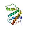 5ijmS S: Starting model for refinement |
|---|---|
| Similar structure data |
-Links
 PDBj
PDBj
- Assembly
| Deposited unit | 
| ||||||||||||
|---|---|---|---|---|---|---|---|---|---|---|---|---|---|
| 1 |
| ||||||||||||
| Unit cell |
|
-Components
| #1: Protein | Mass: 11462.015 Da / Num. of mol.: 1 Source method: isolated from a genetically manipulated source Source: (gene. exp.) Leishmania major (eukaryote) / Gene: LMJF_17_0620 / Production host:  | ||||
|---|---|---|---|---|---|
| #2: Chemical |   Mass: 46.025 Da / Num. of mol.: 3 / Source method: obtained synthetically / Formula: CH2O2 / Feature type: SUBJECT OF INVESTIGATION Mass: 46.025 Da / Num. of mol.: 3 / Source method: obtained synthetically / Formula: CH2O2 / Feature type: SUBJECT OF INVESTIGATION#3: Water | ChemComp-HOH / |  Mass: 18.015 Da / Num. of mol.: 88 / Source method: isolated from a natural source / Formula: H2O Mass: 18.015 Da / Num. of mol.: 88 / Source method: isolated from a natural source / Formula: H2OHas ligand of interest | Y | |
-Experimental details
-Experiment
| Experiment | Method: X-RAY DIFFRACTION / Number of used crystals: 1 |
|---|
- Sample preparation
| Crystal | Density Matthews: 2.16 Å3/Da / Density % sol: 43.01 % / Description: rod-shaped crystals |
|---|---|
| Crystal grow | Temperature: 289.15 K / Method: vapor diffusion, hanging drop Details: 20% (w/v) PEG 3350, 0.2M Magnesium formate dihydrate |
-Data collection
| Diffraction | Mean temperature: 100 K / Ambient temp details: Oxford Cryostream - 700 series / Serial crystal experiment: N |
|---|---|
| Diffraction source | Source: SYNCHROTRON / Site: RRCAT INDUS-2  / Beamline: PX-BL21 / Wavelength: 0.97949 Å / Beamline: PX-BL21 / Wavelength: 0.97949 Å |
| Detector | Type: MAR scanner 345 mm plate / Detector: IMAGE PLATE / Date: Mar 25, 2018 |
| Radiation | Monochromator: Mirrors / Protocol: SINGLE WAVELENGTH / Monochromatic (M) / Laue (L): M / Scattering type: x-ray |
| Radiation wavelength | Wavelength: 0.97949 Å / Relative weight: 1 |
| Reflection | Resolution: 1.4→40.68 Å / Num. obs: 17287 / % possible obs: 99 % / Redundancy: 3.9 % / Biso Wilson estimate: 14.84 Å2 / CC1/2: 0.999 / Rmerge(I) obs: 0.033 / Net I/σ(I): 19.2 |
| Reflection shell | Resolution: 1.4→1.42 Å / Rmerge(I) obs: 0.524 / Mean I/σ(I) obs: 2.2 / Num. unique obs: 978 / % possible all: 99.9 |
- Processing
| Software |
| ||||||||||||||||||||||||||||||||||||||||||||||||||||||||
|---|---|---|---|---|---|---|---|---|---|---|---|---|---|---|---|---|---|---|---|---|---|---|---|---|---|---|---|---|---|---|---|---|---|---|---|---|---|---|---|---|---|---|---|---|---|---|---|---|---|---|---|---|---|---|---|---|---|
| Refinement | Method to determine structure: MOLECULAR REPLACEMENT Starting model: 5IJM Resolution: 1.45→22.77 Å / SU ML: 0.1152 / Cross valid method: FREE R-VALUE / σ(F): 1.38 / Phase error: 19.2643 Stereochemistry target values: GeoStd + Monomer Library + CDL v1.2
| ||||||||||||||||||||||||||||||||||||||||||||||||||||||||
| Solvent computation | Shrinkage radii: 0.9 Å / VDW probe radii: 1.11 Å / Solvent model: FLAT BULK SOLVENT MODEL | ||||||||||||||||||||||||||||||||||||||||||||||||||||||||
| Displacement parameters | Biso mean: 23.04 Å2 | ||||||||||||||||||||||||||||||||||||||||||||||||||||||||
| Refinement step | Cycle: LAST / Resolution: 1.45→22.77 Å
| ||||||||||||||||||||||||||||||||||||||||||||||||||||||||
| Refine LS restraints |
| ||||||||||||||||||||||||||||||||||||||||||||||||||||||||
| LS refinement shell |
|
