 Movie
Movie Controller

[English] 日本語
 Yorodumi
- PDB-7dbs: Crystal Structure Of Biotin Protein Ligase From Leishmania Major ... -
+ Open data

- Basic information
| Entry | Database: PDB / ID: 7dbs | ||||||
|---|---|---|---|---|---|---|---|
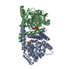
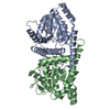
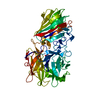
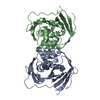
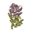
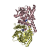
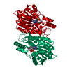
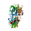
| Title | Crystal Structure Of Biotin Protein Ligase From Leishmania Major in complex with Biotin | ||||||
 Components Components | Biotin/lipoate protein ligase-like protein | ||||||
 Keywords Keywords | LIGASE / Biotin protein ligase / Enzyme | ||||||
| Function / homology |  Function and homology information Function and homology informationbiotin-[biotin carboxyl-carrier protein] ligase / biotin--[biotin carboxyl-carrier protein] ligase activity / cytoplasm Similarity search - Function | ||||||
| Biological species |  Leishmania major (eukaryote) Leishmania major (eukaryote) | ||||||
| Method |  X-RAY DIFFRACTION / SYNCHROTRON / MOLECULAR REPLACEMENT / Resolution: 2.33 Å X-RAY DIFFRACTION / SYNCHROTRON / MOLECULAR REPLACEMENT / Resolution: 2.33 Å | ||||||
 Authors Authors | Rajak, M. / Sundd, M. | ||||||
 Citation Citation | Journal: Acta Crystallogr D Struct Biol / Year: 2021 Title: Leishmania major biotin protein ligase forms a unique cross-handshake dimer. Authors: Rajak, M.K. / Bhatnagar, S. / Pandey, S. / Kumar, S. / Verma, S. / Patel, A.K. / Sundd, M. | ||||||
| History |
|
- Structure visualization
| Structure viewer | Molecule: MolmilJmol/JSmol |
|---|
- Downloads & links
-Download
| PDBx/mmCIF format | 7dbs.cif.gz | 132.2 KB | Display | PDBx/mmCIF format |
|---|---|---|---|---|
| PDB format | pdb7dbs.ent.gz | 84.8 KB | Display | PDB format |
| PDBx/mmJSON format | 7dbs.json.gz | Tree view | PDBx/mmJSON format | |
| Others |  Other downloads Other downloads |
-Validation report
| Arichive directory | https://data.pdbj.org/pub/pdb/validation_reports/db/7dbsftp://data.pdbj.org/pub/pdb/validation_reports/db/7dbs | HTTPS FTP |
|---|
-Related structure data
| Related structure data |  6jhuS S: Starting model for refinement |
|---|---|
| Similar structure data |


-Links
 PDBj
PDBj
- Assembly
| Deposited unit | 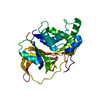
| ||||||||||||
|---|---|---|---|---|---|---|---|---|---|---|---|---|---|
| 1 | 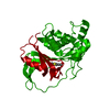
| ||||||||||||
| Unit cell |
|
-Components
| #1: Protein | Mass: 30560.057 Da / Num. of mol.: 1 Source method: isolated from a genetically manipulated source Source: (gene. exp.) Leishmania major (eukaryote) / Gene: LMJF_31_1070 / Production host:  References: UniProt: Q4Q6F6, biotin-[biotin carboxyl-carrier protein] ligase |
|---|---|
| #2: Chemical | ChemComp-BTN /   Mass: 244.311 Da / Num. of mol.: 1 / Source method: obtained synthetically / Formula: C10H16N2O3S / Feature type: SUBJECT OF INVESTIGATION Mass: 244.311 Da / Num. of mol.: 1 / Source method: obtained synthetically / Formula: C10H16N2O3S / Feature type: SUBJECT OF INVESTIGATION |
| #3: Water | ChemComp-HOH /  Mass: 18.015 Da / Num. of mol.: 54 / Source method: isolated from a natural source / Formula: H2O Mass: 18.015 Da / Num. of mol.: 54 / Source method: isolated from a natural source / Formula: H2O |
| Has ligand of interest | Y |
-Experimental details
-Experiment
| Experiment | Method: X-RAY DIFFRACTION / Number of used crystals: 1 |
|---|
- Sample preparation
| Crystal | Density Matthews: 2.31 Å3/Da / Density % sol: 46.9 % |
|---|---|
| Crystal grow | Temperature: 291.15 K / Method: vapor diffusion, hanging drop / Details: 19.5% PEG 3350, 200 mM Ammonium sulfate |
-Data collection
| Diffraction | Mean temperature: 277.15 K / Serial crystal experiment: N |
|---|---|
| Diffraction source | Source: SYNCHROTRON / Site: ESRF  / Beamline: ID29 / Wavelength: 0.979507 Å / Beamline: ID29 / Wavelength: 0.979507 Å |
| Detector | Type: DECTRIS PILATUS3 S 6M / Detector: PIXEL / Date: Jul 23, 2018 |
| Radiation | Protocol: SINGLE WAVELENGTH / Monochromatic (M) / Laue (L): M / Scattering type: x-ray |
| Radiation wavelength | Wavelength: 0.979507 Å / Relative weight: 1 |
| Reflection | Resolution: 2.33→52.84 Å / Num. obs: 11509 / % possible obs: 98.1 % / Redundancy: 6.6 % / Biso Wilson estimate: 64.68 Å2 / CC1/2: 0.996 / Rmerge(I) obs: 0.062 / Rpim(I) all: 0.027 / Net I/σ(I): 13.2 |
| Reflection shell | Resolution: 2.332→2.372 Å / Rmerge(I) obs: 0.631 / Mean I/σ(I) obs: 2.4 / Num. unique obs: 567 / CC1/2: 0.949 / Rpim(I) all: 0.258 / % possible all: 100 |
- Processing
| Software |
| ||||||||||||||||||||||||||||||||||||||||
|---|---|---|---|---|---|---|---|---|---|---|---|---|---|---|---|---|---|---|---|---|---|---|---|---|---|---|---|---|---|---|---|---|---|---|---|---|---|---|---|---|---|
| Refinement | Method to determine structure: MOLECULAR REPLACEMENT Starting model: 6JHU Resolution: 2.33→52.84 Å / SU ML: 0.3804 / Cross valid method: FREE R-VALUE / σ(F): 1.37 / Phase error: 39.9543 Stereochemistry target values: GeoStd + Monomer Library + CDL v1.2
| ||||||||||||||||||||||||||||||||||||||||
| Solvent computation | Shrinkage radii: 0.9 Å / VDW probe radii: 1.11 Å / Solvent model: FLAT BULK SOLVENT MODEL | ||||||||||||||||||||||||||||||||||||||||
| Displacement parameters | Biso mean: 79.59 Å2 | ||||||||||||||||||||||||||||||||||||||||
| Refinement step | Cycle: LAST / Resolution: 2.33→52.84 Å
| ||||||||||||||||||||||||||||||||||||||||
| Refine LS restraints |
| ||||||||||||||||||||||||||||||||||||||||
| LS refinement shell |
| ||||||||||||||||||||||||||||||||||||||||
| Refinement TLS params. | Method: refined / Origin x: 36.135690271 Å / Origin y: 0.685091776768 Å / Origin z: 38.7954802389 Å
| ||||||||||||||||||||||||||||||||||||||||
| Refinement TLS group | Selection details: all |
