 Movie
Movie Controller

[English] 日本語
 Yorodumi
- PDB-7db8: Crystal structure of Mycobacterium tuberculosis phenylalanyl-tRNA... -
+ Open data

- Basic information
| Entry | Database: PDB / ID: 7db8 | ||||||
|---|---|---|---|---|---|---|---|
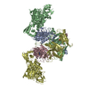
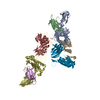
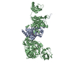
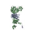
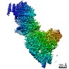
| Title | Crystal structure of Mycobacterium tuberculosis phenylalanyl-tRNA synthetase in complex with compound PF-3845 | ||||||
 Components Components |
| ||||||
 Keywords Keywords | LIGASE / aminoacylation / tRNA-binding / aminoacyl-tRNA synthetase / ATP-binding | ||||||
| Function / homology |  Function and homology information Function and homology informationphenylalanine-tRNA ligase complex / phenylalanine-tRNA ligase / phenylalanyl-tRNA aminoacylation / phenylalanine-tRNA ligase activity / peptidoglycan-based cell wall / tRNA binding / magnesium ion binding / ATP binding / plasma membrane / cytoplasm Similarity search - Function | ||||||
| Biological species |   Mycobacterium tuberculosis (bacteria) Mycobacterium tuberculosis (bacteria) | ||||||
| Method |  X-RAY DIFFRACTION / SYNCHROTRON / MOLECULAR REPLACEMENT / Resolution: 2.3 Å X-RAY DIFFRACTION / SYNCHROTRON / MOLECULAR REPLACEMENT / Resolution: 2.3 Å | ||||||
 Authors Authors | Xu, M. / Zhang, X. / Xu, L. / Chen, S. | ||||||
 Citation Citation | Journal: J.Biol.Chem. / Year: 2021 Title: Re-discovery of PF-3845 as a new chemical scaffold inhibiting phenylalanyl-tRNA synthetase in Mycobacterium tuberculosis . Authors: Wang, H. / Xu, M. / Engelhart, C.A. / Zhang, X. / Yan, B. / Pan, M. / Xu, Y. / Fan, S. / Liu, R. / Xu, L. / Hua, L. / Schnappinger, D. / Chen, S. #1: Journal: J.Biol.Chem. / Year: 2021Title: Re-discovery of PF-3845 as a new chemical scaffold inhibiting phenylalanyl-tRNA synthetase in Mycobacterium tuberculosis Authors: Wang, H. / Xu, M. / Engelhart, C.A. / Zhang, X. / Yan, B. / Pan, M. / Xu, Y. / Fan, S. / Liu, R. / Xu, L. / Hua, L. / Schnappinger, D. / Chen, S. | ||||||
| History |
|
- Structure visualization
| Structure viewer | Molecule: MolmilJmol/JSmol |
|---|
- Downloads & links
-Download
| PDBx/mmCIF format | 7db8.cif.gz | 254.2 KB | Display | PDBx/mmCIF format |
|---|---|---|---|---|
| PDB format | pdb7db8.ent.gz | 197.1 KB | Display | PDB format |
| PDBx/mmJSON format | 7db8.json.gz | Tree view | PDBx/mmJSON format | |
| Others |  Other downloads Other downloads |
-Validation report
| Arichive directory | https://data.pdbj.org/pub/pdb/validation_reports/db/7db8ftp://data.pdbj.org/pub/pdb/validation_reports/db/7db8 | HTTPS FTP |
|---|
-Related structure data
| Related structure data | 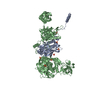 7dawSC  7db7C S: Starting model for refinement C: citing same article ( |
|---|---|
| Similar structure data |


-Links
 PDBj
PDBj

- Assembly
| Deposited unit | 
| ||||||||||||
|---|---|---|---|---|---|---|---|---|---|---|---|---|---|
| 1 | 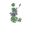
| ||||||||||||
| Unit cell |
|
-Components
| #1: Protein | Mass: 37415.266 Da / Num. of mol.: 1 Source method: isolated from a genetically manipulated source Source: (gene. exp.) Mycobacterium tuberculosis (strain ATCC 25618 / H37Rv) (bacteria)Strain: ATCC 25618 / H37Rv / Gene: pheS, Rv1649, MTCY06H11.14 / Production host: | ||||||
|---|---|---|---|---|---|---|---|
| #2: Protein | Mass: 90179.797 Da / Num. of mol.: 1 Source method: isolated from a genetically manipulated source Source: (gene. exp.) Mycobacterium tuberculosis (strain ATCC 25618 / H37Rv) (bacteria)Strain: ATCC 25618 / H37Rv / Gene: pheT, Rv1650, MTCY06H11.15 / Production host: | ||||||
| #3: Chemical | ChemComp-SO4 /   Mass: 96.063 Da / Num. of mol.: 13 / Source method: obtained synthetically / Formula: SO4 Mass: 96.063 Da / Num. of mol.: 13 / Source method: obtained synthetically / Formula: SO4#4: Chemical | ChemComp-H2R / |   Mass: 456.460 Da / Num. of mol.: 1 / Source method: obtained synthetically / Formula: C24H23F3N4O2 / Feature type: SUBJECT OF INVESTIGATION / Comment: inhibitor*YM Mass: 456.460 Da / Num. of mol.: 1 / Source method: obtained synthetically / Formula: C24H23F3N4O2 / Feature type: SUBJECT OF INVESTIGATION / Comment: inhibitor*YM#5: Water | ChemComp-HOH / |  Mass: 18.015 Da / Num. of mol.: 1026 / Source method: isolated from a natural source / Formula: H2O Mass: 18.015 Da / Num. of mol.: 1026 / Source method: isolated from a natural source / Formula: H2OHas ligand of interest | Y | |
-Experimental details
-Experiment
| Experiment | Method: X-RAY DIFFRACTION / Number of used crystals: 1 |
|---|
- Sample preparation
| Crystal grow | Temperature: 291 K / Method: vapor diffusion, sitting drop / pH: 7.5 / Details: 1.5M (NH4)2SO4, 0.2% PEG 3350, 0.1M HEPES pH 7.5 |
|---|
-Data collection
| Diffraction | Mean temperature: 100 K / Serial crystal experiment: N |
|---|---|
| Diffraction source | Source: SYNCHROTRON / Site: SSRF  / Beamline: BL19U1 / Wavelength: 0.9793 Å / Beamline: BL19U1 / Wavelength: 0.9793 Å |
| Detector | Type: DECTRIS PILATUS3 6M / Detector: PIXEL / Date: Jul 13, 2020 |
| Radiation | Protocol: SINGLE WAVELENGTH / Monochromatic (M) / Laue (L): M / Scattering type: x-ray |
| Radiation wavelength | Wavelength: 0.9793 Å / Relative weight: 1 |
| Reflection | Resolution: 2.3→50 Å / Num. obs: 121928 / % possible obs: 99.9 % / Redundancy: 13.4 % / Biso Wilson estimate: 29.83 Å2 / CC1/2: 0.994 / Rmerge(I) obs: 0.138 / Net I/σ(I): 19 |
| Reflection shell | Resolution: 2.3→2.34 Å / Redundancy: 13.7 % / Rmerge(I) obs: 0.681 / Mean I/σ(I) obs: 4.1 / Num. unique obs: 6050 / CC1/2: 0.953 / % possible all: 100 |
- Processing
| Software |
| |||||||||||||||||||||||||||||||||||||||||||||||||||||||||||||||||||||||||||||||||||||||||||||||||||||||||||||||||||||||||||||||||||||||||||||||||||||||||||||||||||||||||||||||||||||||||||||||||||||||||||||||||||||||||
|---|---|---|---|---|---|---|---|---|---|---|---|---|---|---|---|---|---|---|---|---|---|---|---|---|---|---|---|---|---|---|---|---|---|---|---|---|---|---|---|---|---|---|---|---|---|---|---|---|---|---|---|---|---|---|---|---|---|---|---|---|---|---|---|---|---|---|---|---|---|---|---|---|---|---|---|---|---|---|---|---|---|---|---|---|---|---|---|---|---|---|---|---|---|---|---|---|---|---|---|---|---|---|---|---|---|---|---|---|---|---|---|---|---|---|---|---|---|---|---|---|---|---|---|---|---|---|---|---|---|---|---|---|---|---|---|---|---|---|---|---|---|---|---|---|---|---|---|---|---|---|---|---|---|---|---|---|---|---|---|---|---|---|---|---|---|---|---|---|---|---|---|---|---|---|---|---|---|---|---|---|---|---|---|---|---|---|---|---|---|---|---|---|---|---|---|---|---|---|---|---|---|---|---|---|---|---|---|---|---|---|---|---|---|---|---|---|---|---|
| Refinement | Method to determine structure: MOLECULAR REPLACEMENT Starting model: 7DAW Resolution: 2.3→49.64 Å / SU ML: 0.1896 / Cross valid method: FREE R-VALUE / σ(F): 1.36 / Phase error: 18.7042 Stereochemistry target values: GeoStd + Monomer Library + CDL v1.2
| |||||||||||||||||||||||||||||||||||||||||||||||||||||||||||||||||||||||||||||||||||||||||||||||||||||||||||||||||||||||||||||||||||||||||||||||||||||||||||||||||||||||||||||||||||||||||||||||||||||||||||||||||||||||||
| Solvent computation | Shrinkage radii: 0.9 Å / VDW probe radii: 1.11 Å / Solvent model: FLAT BULK SOLVENT MODEL | |||||||||||||||||||||||||||||||||||||||||||||||||||||||||||||||||||||||||||||||||||||||||||||||||||||||||||||||||||||||||||||||||||||||||||||||||||||||||||||||||||||||||||||||||||||||||||||||||||||||||||||||||||||||||
| Displacement parameters | Biso mean: 35.25 Å2 | |||||||||||||||||||||||||||||||||||||||||||||||||||||||||||||||||||||||||||||||||||||||||||||||||||||||||||||||||||||||||||||||||||||||||||||||||||||||||||||||||||||||||||||||||||||||||||||||||||||||||||||||||||||||||
| Refinement step | Cycle: LAST / Resolution: 2.3→49.64 Å
| |||||||||||||||||||||||||||||||||||||||||||||||||||||||||||||||||||||||||||||||||||||||||||||||||||||||||||||||||||||||||||||||||||||||||||||||||||||||||||||||||||||||||||||||||||||||||||||||||||||||||||||||||||||||||
| Refine LS restraints |
| |||||||||||||||||||||||||||||||||||||||||||||||||||||||||||||||||||||||||||||||||||||||||||||||||||||||||||||||||||||||||||||||||||||||||||||||||||||||||||||||||||||||||||||||||||||||||||||||||||||||||||||||||||||||||
| LS refinement shell |
|
