 Movie
Movie Controller


+ Open data

- Basic information
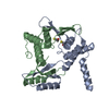
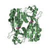
| Entry | Database: PDB / ID: 7d55 | ||||||
|---|---|---|---|---|---|---|---|
| Title | Crystal structure of lys170 CBD | ||||||
 Components Components | Putative N-acetylmuramoyl-L-alanine amidase | ||||||
 Keywords Keywords | HYDROLASE / lysin | ||||||
| Function / homology |  Function and homology information Function and homology informationN-acetylmuramoyl-L-alanine amidase / N-acetylmuramoyl-L-alanine amidase activity / peptidoglycan turnover / peptidoglycan catabolic process / cell wall organization / killing of cells of another organism / defense response to bacterium Similarity search - Function | ||||||
| Biological species |  Enterococcus phage phiM1EF22 (virus) Enterococcus phage phiM1EF22 (virus) | ||||||
| Method |  X-RAY DIFFRACTION / SYNCHROTRON / SAD / Resolution: 1.397 Å X-RAY DIFFRACTION / SYNCHROTRON / SAD / Resolution: 1.397 Å | ||||||
 Authors Authors | Zhen, X. | ||||||
 Citation Citation | Journal: Eur.Biophys.J. / Year: 2021 Title: Structural and biochemical analyses of the tetrameric cell binding domain of Lys170 from enterococcal phage F170/08. Authors: Xu, X. / Zhang, D. / Zhou, B. / Zhen, X. / Ouyang, S. | ||||||
| History |
|
- Structure visualization
| Structure viewer | Molecule: MolmilJmol/JSmol |
|---|
- Downloads & links
-Download
| PDBx/mmCIF format | 7d55.cif.gz | 158 KB | Display | PDBx/mmCIF format |
|---|---|---|---|---|
| PDB format | pdb7d55.ent.gz | 126 KB | Display | PDB format |
| PDBx/mmJSON format | 7d55.json.gz | Tree view | PDBx/mmJSON format | |
| Others |  Other downloads Other downloads |
-Validation report
| Summary document | 7d55_validation.pdf.gz | 429.9 KB | Display | wwPDB validaton report |
|---|---|---|---|---|
| Full document | 7d55_full_validation.pdf.gz | 430.7 KB | Display | |
| Data in XML | 7d55_validation.xml.gz | 18.5 KB | Display | |
| Data in CIF | 7d55_validation.cif.gz | 27.2 KB | Display | |
| Arichive directory | https://data.pdbj.org/pub/pdb/validation_reports/d5/7d55ftp://data.pdbj.org/pub/pdb/validation_reports/d5/7d55 | HTTPS FTP |
-Related structure data
| Similar structure data |
|---|


-Links
 PDBj
PDBj- Assembly
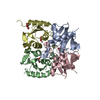
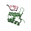
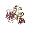
| Deposited unit | 
| ||||||||
|---|---|---|---|---|---|---|---|---|---|
| 1 |
| ||||||||
| Unit cell |
| ||||||||
| Components on special symmetry positions |
|
-Components
| #1: Protein | Mass: 10390.896 Da / Num. of mol.: 4 Source method: isolated from a genetically manipulated source Source: (gene. exp.) Enterococcus phage phiM1EF22 (virus) / Gene: PHIM1EF22_0110 / Production host:  #2: Water | ChemComp-HOH / |  Mass: 18.015 Da / Num. of mol.: 399 / Source method: isolated from a natural source / Formula: H2O Mass: 18.015 Da / Num. of mol.: 399 / Source method: isolated from a natural source / Formula: H2O |
|---|
-Experimental details
-Experiment
| Experiment | Method: X-RAY DIFFRACTION / Number of used crystals: 1 |
|---|
- Sample preparation
| Crystal | Density Matthews: 2.51 Å3/Da / Density % sol: 51.03 % |
|---|---|
| Crystal grow | Temperature: 289 K / Method: vapor diffusion, sitting drop / pH: 6.5 Details: 0.1 M MES pH 6.5, 25% (w/v) PEG 5000 and 1% (w/v) benzamidine hydrochloride PH range: 6.5-9.0 |
-Data collection
| Diffraction | Mean temperature: 100 K / Serial crystal experiment: N |
|---|---|
| Diffraction source | Source: SYNCHROTRON / Site: SSRF  / Beamline: BL17U1 / Wavelength: 0.9791 Å / Beamline: BL17U1 / Wavelength: 0.9791 Å |
| Detector | Type: ADSC QUANTUM 315r / Detector: CCD / Date: Jun 12, 2019 |
| Radiation | Protocol: SINGLE WAVELENGTH / Monochromatic (M) / Laue (L): M / Scattering type: x-ray |
| Radiation wavelength | Wavelength: 0.9791 Å / Relative weight: 1 |
| Reflection | Resolution: 1.397→73.9411 Å / Num. obs: 58593 / % possible obs: 100 % / Redundancy: 12.4 % / CC1/2: 0.998 / Net I/σ(I): 2.7 |
| Reflection shell | Resolution: 1.4→1.45 Å / Num. unique obs: 573 / CC1/2: 0.909 |
- Processing
| Software |
| ||||||||||||||||||||||||||||||||||||||||||||||||||||||||||||||||||||||||||||||||||||||||||
|---|---|---|---|---|---|---|---|---|---|---|---|---|---|---|---|---|---|---|---|---|---|---|---|---|---|---|---|---|---|---|---|---|---|---|---|---|---|---|---|---|---|---|---|---|---|---|---|---|---|---|---|---|---|---|---|---|---|---|---|---|---|---|---|---|---|---|---|---|---|---|---|---|---|---|---|---|---|---|---|---|---|---|---|---|---|---|---|---|---|---|---|
| Refinement | Method to determine structure: SAD / Resolution: 1.397→73.941 Å / SU ML: 0.13 / Cross valid method: THROUGHOUT / σ(F): 1.35 / Phase error: 23.21 / Stereochemistry target values: ML
| ||||||||||||||||||||||||||||||||||||||||||||||||||||||||||||||||||||||||||||||||||||||||||
| Solvent computation | Shrinkage radii: 0.9 Å / VDW probe radii: 1.11 Å / Solvent model: FLAT BULK SOLVENT MODEL | ||||||||||||||||||||||||||||||||||||||||||||||||||||||||||||||||||||||||||||||||||||||||||
| Displacement parameters | Biso max: 72.2 Å2 / Biso mean: 18.8309 Å2 / Biso min: 4.46 Å2 | ||||||||||||||||||||||||||||||||||||||||||||||||||||||||||||||||||||||||||||||||||||||||||
| Refinement step | Cycle: final / Resolution: 1.397→73.941 Å
| ||||||||||||||||||||||||||||||||||||||||||||||||||||||||||||||||||||||||||||||||||||||||||
| Refine LS restraints |
| ||||||||||||||||||||||||||||||||||||||||||||||||||||||||||||||||||||||||||||||||||||||||||
| LS refinement shell | Refine-ID: X-RAY DIFFRACTION / Rfactor Rfree error: 0
|
