 Movie
Movie Controller

[English] 日本語
 Yorodumi
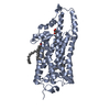
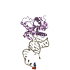
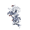
- PDB-7d2z: Structure of sybody SR31 in complex with the SARS-CoV-2 S Recepto... -
+ Open data

- Basic information
| Entry | Database: PDB / ID: 7d2z | |||||||||
|---|---|---|---|---|---|---|---|---|---|---|
| Title | Structure of sybody SR31 in complex with the SARS-CoV-2 S Receptor Binding domain (RBD) | |||||||||
 Components Components |
| |||||||||
 Keywords Keywords | PROTEIN BINDING / coronavirus / Covid-19 / nanobody / neutralizing antibody / receptor binding protein / SARS-CoV-2 / S protein / synthetic antibody / VHH. | |||||||||
| Function / homology |  Function and homology information Function and homology informationsymbiont-mediated disruption of host tissue / Maturation of spike protein / Translation of Structural Proteins / Virion Assembly and Release / host cell surface / host extracellular region / symbiont-mediated-mediated suppression of host tetherin activity / Induction of Cell-Cell Fusion / structural constituent of virion / positive regulation of viral entry into host cell ...symbiont-mediated disruption of host tissue / Maturation of spike protein / Translation of Structural Proteins / Virion Assembly and Release / host cell surface / host extracellular region / symbiont-mediated-mediated suppression of host tetherin activity / Induction of Cell-Cell Fusion / structural constituent of virion / positive regulation of viral entry into host cell / membrane fusion / host cell endoplasmic reticulum-Golgi intermediate compartment membrane / Attachment and Entry / entry receptor-mediated virion attachment to host cell / receptor-mediated virion attachment to host cell / host cell surface receptor binding / symbiont-mediated suppression of host innate immune response / endocytosis involved in viral entry into host cell / receptor ligand activity / fusion of virus membrane with host plasma membrane / fusion of virus membrane with host endosome membrane / viral envelope / symbiont entry into host cell / virion attachment to host cell / host cell plasma membrane / SARS-CoV-2 activates/modulates innate and adaptive immune responses / virion membrane / membrane / identical protein binding / plasma membrane Similarity search - Function | |||||||||
| Biological species | synthetic construct (others)  Severe acute respiratory syndrome coronavirus 2 Severe acute respiratory syndrome coronavirus 2 | |||||||||
| Method |  X-RAY DIFFRACTION / SYNCHROTRON / MOLECULAR REPLACEMENT / Resolution: 1.97 Å X-RAY DIFFRACTION / SYNCHROTRON / MOLECULAR REPLACEMENT / Resolution: 1.97 Å | |||||||||
 Authors Authors | Li, T. / Cai, H. / Yao, H. / Qin, W. / Li, D. | |||||||||
| Funding support |  China, 2items China, 2items
| |||||||||
 Citation Citation | Journal: Plos Pathog. / Year: 2021 Title: A high-affinity RBD-targeting nanobody improves fusion partner's potency against SARS-CoV-2. Authors: Yao, H. / Cai, H. / Li, T. / Zhou, B. / Qin, W. / Lavillette, D. / Li, D. | |||||||||
| History |
|
- Structure visualization
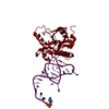
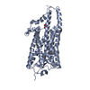
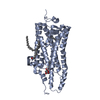
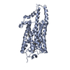
| Structure viewer | Molecule: MolmilJmol/JSmol |
|---|
- Downloads & links
-Download
| PDBx/mmCIF format | 7d2z.cif.gz | 108.3 KB | Display | PDBx/mmCIF format |
|---|---|---|---|---|
| PDB format | pdb7d2z.ent.gz | 65.3 KB | Display | PDB format |
| PDBx/mmJSON format | 7d2z.json.gz | Tree view | PDBx/mmJSON format | |
| Others |  Other downloads Other downloads |
-Validation report
| Arichive directory | https://data.pdbj.org/pub/pdb/validation_reports/d2/7d2zftp://data.pdbj.org/pub/pdb/validation_reports/d2/7d2z | HTTPS FTP |
|---|
-Related structure data
| Related structure data |  7d30C 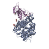 6m0jS S: Starting model for refinement C: citing same article ( |
|---|---|
| Similar structure data |


-Links
 PDBj
PDBj


- Assembly
| Deposited unit | 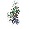
| ||||||||||||
|---|---|---|---|---|---|---|---|---|---|---|---|---|---|
| 1 |
| ||||||||||||
| Unit cell |
|
-Components
-Antibody / Protein / Sugars , 3 types, 3 molecules AB
| #1: Antibody | Mass: 15722.449 Da / Num. of mol.: 1 Source method: isolated from a genetically manipulated source Source: (gene. exp.) synthetic construct (others) / Production host:  |
|---|---|
| #2: Protein | Mass: 23747.643 Da / Num. of mol.: 1 / Fragment: Receptor binding domain (RBD) Source method: isolated from a genetically manipulated source Source: (gene. exp.) Severe acute respiratory syndrome coronavirus 2Gene: S, 2 / Production host:  Trichoplusia ni (cabbage looper) / Strain (production host): Hi5 / References: UniProt: P0DTC2 Trichoplusia ni (cabbage looper) / Strain (production host): Hi5 / References: UniProt: P0DTC2 |
| #3: Polysaccharide | alpha-D-mannopyranose-(1-3)-[alpha-D-mannopyranose-(1-6)]beta-D-mannopyranose-(1-4)-2-acetamido-2- ...alpha-D-mannopyranose-(1-3)-[alpha-D-mannopyranose-(1-6)]beta-D-mannopyranose-(1-4)-2-acetamido-2-deoxy-beta-D-glucopyranose-(1-4)-[alpha-L-fucopyranose-(1-3)][alpha-L-fucopyranose-(1-6)]2-acetamido-2-deoxy-beta-D-glucopyranose Source method: isolated from a genetically manipulated source |
-Non-polymers , 4 types, 186 molecules 


| #4: Chemical | ChemComp-FMT /  Mass: 46.025 Da / Num. of mol.: 13 / Source method: obtained synthetically / Formula: CH2O2 Mass: 46.025 Da / Num. of mol.: 13 / Source method: obtained synthetically / Formula: CH2O2#5: Chemical | ChemComp-GOL /  Mass: 92.094 Da / Num. of mol.: 5 / Source method: obtained synthetically / Formula: C3H8O3 Mass: 92.094 Da / Num. of mol.: 5 / Source method: obtained synthetically / Formula: C3H8O3#6: Chemical |  Mass: 59.044 Da / Num. of mol.: 2 / Source method: obtained synthetically / Formula: C2H3O2 Mass: 59.044 Da / Num. of mol.: 2 / Source method: obtained synthetically / Formula: C2H3O2#7: Water | ChemComp-HOH / | Mass: 18.015 Da / Num. of mol.: 166 / Source method: isolated from a natural source / Formula: H2O |
|---|
-Details
| Has ligand of interest | Y |
|---|---|
| Has protein modification | Y |
-Experimental details
-Experiment
| Experiment | Method: X-RAY DIFFRACTION / Number of used crystals: 1 |
|---|
- Sample preparation
| Crystal | Density Matthews: 3.38 Å3/Da / Density % sol: 63.63 % |
|---|---|
| Crystal grow | Temperature: 293 K / Method: vapor diffusion, sitting drop Details: 2.0M sodium formate, 0.1M sodium acetate trihydrate pH 4.6 |
-Data collection
| Diffraction | Mean temperature: 100 K / Serial crystal experiment: N |
|---|---|
| Diffraction source | Source: SYNCHROTRON / Site: SSRF / Beamline: BL19U1 / Wavelength: 0.97853 Å |
| Detector | Type: PSI PILATUS 6M / Detector: PIXEL / Date: May 6, 2020 |
| Radiation | Protocol: SINGLE WAVELENGTH / Monochromatic (M) / Laue (L): M / Scattering type: x-ray |
| Radiation wavelength | Wavelength: 0.97853 Å / Relative weight: 1 |
| Reflection | Resolution: 1.97→19.62 Å / Num. obs: 35708 / % possible obs: 99.85 % / Redundancy: 19.8 % / Biso Wilson estimate: 42.58 Å2 / CC1/2: 0.999 / Rmerge(I) obs: 0.0907 / Rpim(I) all: 0.02094 / Net I/σ(I): 19.48 |
| Reflection shell | Resolution: 1.97→2.04 Å / Redundancy: 18.8 % / Mean I/σ(I) obs: 1.67 / Num. unique obs: 3516 / CC1/2: 0.819 / CC star: 0.949 / Rpim(I) all: 0.3355 / Rsym value: 1.425 / % possible all: 99.97 |
- Processing
| Software |
| |||||||||||||||||||||||||||||||||||||||||||||||||||||||||||||||||||||||||||||||||||||||||||
|---|---|---|---|---|---|---|---|---|---|---|---|---|---|---|---|---|---|---|---|---|---|---|---|---|---|---|---|---|---|---|---|---|---|---|---|---|---|---|---|---|---|---|---|---|---|---|---|---|---|---|---|---|---|---|---|---|---|---|---|---|---|---|---|---|---|---|---|---|---|---|---|---|---|---|---|---|---|---|---|---|---|---|---|---|---|---|---|---|---|---|---|---|
| Refinement | Method to determine structure: MOLECULAR REPLACEMENT Starting model: 6M0J Resolution: 1.97→19.62 Å / SU ML: 0.2057 / Cross valid method: FREE R-VALUE / σ(F): 1.34 / Phase error: 23.0954 Stereochemistry target values: GeoStd + Monomer Library + CDL v1.2
| |||||||||||||||||||||||||||||||||||||||||||||||||||||||||||||||||||||||||||||||||||||||||||
| Solvent computation | Shrinkage radii: 0.9 Å / VDW probe radii: 1.11 Å / Solvent model: FLAT BULK SOLVENT MODEL | |||||||||||||||||||||||||||||||||||||||||||||||||||||||||||||||||||||||||||||||||||||||||||
| Displacement parameters | Biso mean: 49.49 Å2 | |||||||||||||||||||||||||||||||||||||||||||||||||||||||||||||||||||||||||||||||||||||||||||
| Refinement step | Cycle: LAST / Resolution: 1.97→19.62 Å
| |||||||||||||||||||||||||||||||||||||||||||||||||||||||||||||||||||||||||||||||||||||||||||
| Refine LS restraints |
| |||||||||||||||||||||||||||||||||||||||||||||||||||||||||||||||||||||||||||||||||||||||||||
| LS refinement shell |
|
