 Movie
Movie Controller


+ Open data

- Basic information
| Entry | Database: PDB / ID: 7d04 | ||||||||||||
|---|---|---|---|---|---|---|---|---|---|---|---|---|---|
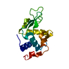
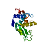
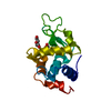
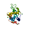
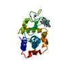
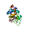
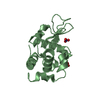
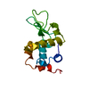
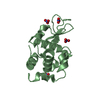
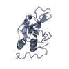
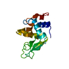
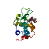
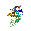
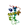
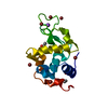
| Title | Lysozyme structure SS3 from SS mode | ||||||||||||
 Components Components | Lysozyme C | ||||||||||||
 Keywords Keywords | HYDROLASE / lysozyme / XFEL / SFX / SElf-seeded mode | ||||||||||||
| Function / homology |  Function and homology information Function and homology informationLactose synthesis / Antimicrobial peptides / Neutrophil degranulation / beta-N-acetylglucosaminidase activity / cell wall macromolecule catabolic process / lysozyme / lysozyme activity / killing of cells of another organism / defense response to Gram-negative bacterium / defense response to bacterium ...Lactose synthesis / Antimicrobial peptides / Neutrophil degranulation / beta-N-acetylglucosaminidase activity / cell wall macromolecule catabolic process / lysozyme / lysozyme activity / killing of cells of another organism / defense response to Gram-negative bacterium / defense response to bacterium / defense response to Gram-positive bacterium / endoplasmic reticulum / : / identical protein binding / cytoplasm Similarity search - Function | ||||||||||||
| Biological species |  | ||||||||||||
| Method |  X-RAY DIFFRACTION / FREE ELECTRON LASER / MOLECULAR REPLACEMENT / Resolution: 1.7 Å X-RAY DIFFRACTION / FREE ELECTRON LASER / MOLECULAR REPLACEMENT / Resolution: 1.7 Å | ||||||||||||
 Authors Authors | Kang, H.S. / Lee, S.J. | ||||||||||||
| Funding support |  Korea, Republic Of, 3items Korea, Republic Of, 3items
| ||||||||||||
 Citation Citation | Journal: Nat Photonics / Year: 2021 Title: High-brightness self-seeded X-ray free-electron laser covering the 3.5 keV to 14.6 keV range Authors: Nam, I. / Min, C.K. / Oh, B. / Kim, G. / Na, D. / Suh, Y.J. / Yang, H. / Cho, M.H. / Kim, C. / Kim, M.J. / Shim, C.H. / Ko, J.H. / Heo, H. / Park, J. / Kim, J. / Park, S. / Park, G. / Kim, S. ...Authors: Nam, I. / Min, C.K. / Oh, B. / Kim, G. / Na, D. / Suh, Y.J. / Yang, H. / Cho, M.H. / Kim, C. / Kim, M.J. / Shim, C.H. / Ko, J.H. / Heo, H. / Park, J. / Kim, J. / Park, S. / Park, G. / Kim, S. / Chun, S.H. / Hyun, H. / Lee, J.H. / Kim, K.S. / Eom, I. / Rah, S. / Shu, D. / Kim, K.J. / Terentyev, S. / Blank, V. / Shvydko, Y. / Lee, S.J. / Kang, H.S. | ||||||||||||
| History |
|
- Structure visualization
| Structure viewer | Molecule: MolmilJmol/JSmol |
|---|
- Downloads & links
-Download
| PDBx/mmCIF format | 7d04.cif.gz | 40.9 KB | Display | PDBx/mmCIF format |
|---|---|---|---|---|
| PDB format | pdb7d04.ent.gz | 26.3 KB | Display | PDB format |
| PDBx/mmJSON format | 7d04.json.gz | Tree view | PDBx/mmJSON format | |
| Others |  Other downloads Other downloads |
-Validation report
| Arichive directory | https://data.pdbj.org/pub/pdb/validation_reports/d0/7d04ftp://data.pdbj.org/pub/pdb/validation_reports/d0/7d04 | HTTPS FTP |
|---|
-Related structure data
| Related structure data | 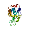 7byoC 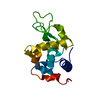 7bypC 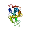 7d01C  7d02C 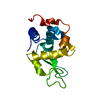 7d05C 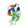 1vdsS S: Starting model for refinement C: citing same article ( |
|---|---|
| Similar structure data |
-Links
 PDBj
PDBj


- Assembly
| Deposited unit | 
| ||||||||||||
|---|---|---|---|---|---|---|---|---|---|---|---|---|---|
| 1 |
| ||||||||||||
| Unit cell |
| ||||||||||||
| Components on special symmetry positions |
|
-Components
| #1: Protein | Mass: 16257.660 Da / Num. of mol.: 1 Source method: isolated from a genetically manipulated source Source: (gene. exp.)  |
|---|---|
| #2: Water | ChemComp-HOH /  Mass: 18.015 Da / Num. of mol.: 72 / Source method: isolated from a natural source / Formula: H2O Mass: 18.015 Da / Num. of mol.: 72 / Source method: isolated from a natural source / Formula: H2O |
| Has protein modification | Y |
-Experimental details
-Experiment
| Experiment | Method: X-RAY DIFFRACTION / Number of used crystals: 1 |
|---|
- Sample preparation
| Crystal | Density Matthews: 1.97 Å3/Da / Density % sol: 32.03 % Description: THE ENTRY CONTAINS FRIEDEL PAIRS IN I_PLUS/MINUS COLUMNS. |
|---|---|
| Crystal grow | Temperature: 293 K / Method: batch mode Details: 100 mM sodium acetate (pH 4.0), 6% (w/v) polyethylene glycol 8,000, and 3.5 M NaCl |
-Data collection
| Diffraction | Mean temperature: 293 K / Serial crystal experiment: Y |
|---|---|
| Diffraction source | Source: FREE ELECTRON LASER / Site: PAL-XFEL / Beamline: NCI / Wavelength: 1.2782 Å |
| Detector | Type: RAYONIX MX-225 / Detector: CCD / Date: Nov 28, 2019 |
| Radiation | Protocol: SINGLE WAVELENGTH / Monochromatic (M) / Laue (L): M / Scattering type: x-ray |
| Radiation wavelength | Wavelength: 1.2782 Å / Relative weight: 1 |
| Reflection | Resolution: 1.7→27.89 Å / Num. obs: 25173 / % possible obs: 100 % / Redundancy: 140.6 % / CC star: 0.964 / R split: 0.295 / Net I/σ(I): 3.52 |
| Reflection shell | Resolution: 1.7→1.73 Å / Mean I/σ(I) obs: 1.56 / Num. unique obs: 2532 / CC star: 0.869 / R split: 0.647 |
| Serial crystallography sample delivery | Method: injection |
- Processing
| Software |
| ||||||||||||||||||||||||||||||||||||||||||||||||||||||||||||||||||||||
|---|---|---|---|---|---|---|---|---|---|---|---|---|---|---|---|---|---|---|---|---|---|---|---|---|---|---|---|---|---|---|---|---|---|---|---|---|---|---|---|---|---|---|---|---|---|---|---|---|---|---|---|---|---|---|---|---|---|---|---|---|---|---|---|---|---|---|---|---|---|---|---|
| Refinement | Method to determine structure: MOLECULAR REPLACEMENT Starting model: 1VDS Resolution: 1.7→27.89 Å / SU ML: 0.22 / Cross valid method: FREE R-VALUE / σ(F): 1.35 / Phase error: 25.67 / Stereochemistry target values: ML
| ||||||||||||||||||||||||||||||||||||||||||||||||||||||||||||||||||||||
| Solvent computation | Shrinkage radii: 0.9 Å / VDW probe radii: 1.11 Å / Solvent model: FLAT BULK SOLVENT MODEL | ||||||||||||||||||||||||||||||||||||||||||||||||||||||||||||||||||||||
| Refinement step | Cycle: LAST / Resolution: 1.7→27.89 Å
| ||||||||||||||||||||||||||||||||||||||||||||||||||||||||||||||||||||||
| Refine LS restraints |
| ||||||||||||||||||||||||||||||||||||||||||||||||||||||||||||||||||||||
| LS refinement shell |
|
