 Movie
Movie Controller


+ Open data

- Basic information
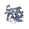
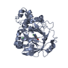
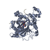
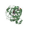
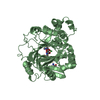
| Entry | Database: PDB / ID: 7cv8 | ||||||
|---|---|---|---|---|---|---|---|
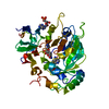
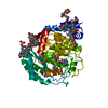
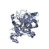
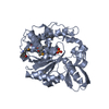
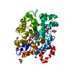
| Title | RNA methyltransferase METTL4 | ||||||
 Components Components | Methyltransferase-like protein 2 | ||||||
 Keywords Keywords | NUCLEAR PROTEIN / RNA methyltransferase | ||||||
| Function / homology |  Function and homology information Function and homology informationsnRNA methyltransferase activity / heat acclimation / DNA-methyltransferase activity / Transferases; Transferring one-carbon groups; Methyltransferases / methyltransferase activity / methylation / nucleic acid binding Similarity search - Function | ||||||
| Biological species |  | ||||||
| Method |  X-RAY DIFFRACTION / SYNCHROTRON / SAD / Resolution: 2.37 Å X-RAY DIFFRACTION / SYNCHROTRON / SAD / Resolution: 2.37 Å | ||||||
 Authors Authors | Luo, Q. / Ma, J. | ||||||
 Citation Citation | Journal: Nat Commun / Year: 2022 Title: Structural insights into molecular mechanism for N6-adenosine methylation by MT-A70 family methyltransferase METTL4 Authors: Luo, Q. / Mo, J. / Chen, H. / Hu, Z. / Wang, B. / Wu, J. / Liang, Z. / Xie, W. / Du, K. / Peng, M. / Li, Y. / Li, T. / Zhang, Y. / Shi, X. / Shen, W.H. / Shi, Y. / Dong, A. / Wang, H. / Ma, J. | ||||||
| History |
|
- Structure visualization
| Structure viewer | Molecule: MolmilJmol/JSmol |
|---|
- Downloads & links
-Download
| PDBx/mmCIF format | 7cv8.cif.gz | 156.4 KB | Display | PDBx/mmCIF format |
|---|---|---|---|---|
| PDB format | pdb7cv8.ent.gz | 121.8 KB | Display | PDB format |
| PDBx/mmJSON format | 7cv8.json.gz | Tree view | PDBx/mmJSON format | |
| Others |  Other downloads Other downloads |
-Validation report
| Arichive directory | https://data.pdbj.org/pub/pdb/validation_reports/cv/7cv8ftp://data.pdbj.org/pub/pdb/validation_reports/cv/7cv8 | HTTPS FTP |
|---|
-Related structure data

-Links
 PDBj
PDBj
- Assembly
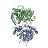
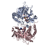
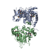
| Deposited unit | 
| ||||||||
|---|---|---|---|---|---|---|---|---|---|
| 1 |
| ||||||||
| Unit cell |
|
-Components
| #1: Protein | Mass: 47955.102 Da / Num. of mol.: 1 Source method: isolated from a genetically manipulated source Source: (gene. exp.)  References: UniProt: Q8LFA9, Transferases; Transferring one-carbon groups; Methyltransferases | ||||
|---|---|---|---|---|---|
| #2: Chemical | ChemComp-SFG /   Mass: 381.387 Da / Num. of mol.: 1 / Source method: obtained synthetically / Formula: C15H23N7O5 / Feature type: SUBJECT OF INVESTIGATION Mass: 381.387 Da / Num. of mol.: 1 / Source method: obtained synthetically / Formula: C15H23N7O5 / Feature type: SUBJECT OF INVESTIGATION | ||||
| #3: Chemical |   Mass: 92.094 Da / Num. of mol.: 3 / Source method: obtained synthetically / Formula: C3H8O3 Mass: 92.094 Da / Num. of mol.: 3 / Source method: obtained synthetically / Formula: C3H8O3#4: Water | ChemComp-HOH / |  Mass: 18.015 Da / Num. of mol.: 61 / Source method: isolated from a natural source / Formula: H2O Mass: 18.015 Da / Num. of mol.: 61 / Source method: isolated from a natural source / Formula: H2OHas ligand of interest | Y | |
-Experimental details
-Experiment
| Experiment | Method: X-RAY DIFFRACTION / Number of used crystals: 1 |
|---|
- Sample preparation
| Crystal | Density Matthews: 2.17 Å3/Da / Density % sol: 43.23 % |
|---|---|
| Crystal grow | Temperature: 291 K / Method: vapor diffusion, sitting drop / Details: 1M Lithium chloride 0.1M HEPES pH 7.0 10% PEG6000 |
-Data collection
| Diffraction | Mean temperature: 100 K / Serial crystal experiment: N | |||||||||||||||||||||||||||||||||||||||||||||||||||||||||||||||||||||||||||||||||||||||||||||||||||
|---|---|---|---|---|---|---|---|---|---|---|---|---|---|---|---|---|---|---|---|---|---|---|---|---|---|---|---|---|---|---|---|---|---|---|---|---|---|---|---|---|---|---|---|---|---|---|---|---|---|---|---|---|---|---|---|---|---|---|---|---|---|---|---|---|---|---|---|---|---|---|---|---|---|---|---|---|---|---|---|---|---|---|---|---|---|---|---|---|---|---|---|---|---|---|---|---|---|---|---|---|
| Diffraction source | Source: SYNCHROTRON / Site: SSRF  / Beamline: BL19U1 / Wavelength: 0.97776 Å / Beamline: BL19U1 / Wavelength: 0.97776 Å | |||||||||||||||||||||||||||||||||||||||||||||||||||||||||||||||||||||||||||||||||||||||||||||||||||
| Detector | Type: DECTRIS EIGER X 16M / Detector: PIXEL / Date: Jun 11, 2016 | |||||||||||||||||||||||||||||||||||||||||||||||||||||||||||||||||||||||||||||||||||||||||||||||||||
| Radiation | Protocol: SINGLE WAVELENGTH / Monochromatic (M) / Laue (L): M / Scattering type: x-ray | |||||||||||||||||||||||||||||||||||||||||||||||||||||||||||||||||||||||||||||||||||||||||||||||||||
| Radiation wavelength | Wavelength: 0.97776 Å / Relative weight: 1 | |||||||||||||||||||||||||||||||||||||||||||||||||||||||||||||||||||||||||||||||||||||||||||||||||||
| Reflection | Resolution: 2.35→46.96 Å / Num. obs: 17743 / % possible obs: 98.8 % / Redundancy: 11.8 % / Rmerge(I) obs: 0.18 / Rpim(I) all: 0.054 / Rrim(I) all: 0.188 / Χ2: 1.95 / Net I/σ(I): 4.7 | |||||||||||||||||||||||||||||||||||||||||||||||||||||||||||||||||||||||||||||||||||||||||||||||||||
| Reflection shell | Diffraction-ID: 1
|
- Processing
| Software |
| ||||||||||||||||||||||||||||||||||||||||||||||||||||||||||||
|---|---|---|---|---|---|---|---|---|---|---|---|---|---|---|---|---|---|---|---|---|---|---|---|---|---|---|---|---|---|---|---|---|---|---|---|---|---|---|---|---|---|---|---|---|---|---|---|---|---|---|---|---|---|---|---|---|---|---|---|---|---|
| Refinement | Method to determine structure: SAD / Resolution: 2.37→30 Å / Cor.coef. Fo:Fc: 0.952 / Cor.coef. Fo:Fc free: 0.924 / SU B: 10.389 / SU ML: 0.132 / Cross valid method: THROUGHOUT / σ(F): 0 / ESU R: 0.085 / ESU R Free: 0.059 / Stereochemistry target values: MAXIMUM LIKELIHOOD Details: HYDROGENS HAVE BEEN ADDED IN THE RIDING POSITIONS U VALUES : WITH TLS ADDED
| ||||||||||||||||||||||||||||||||||||||||||||||||||||||||||||
| Solvent computation | Ion probe radii: 0.8 Å / Shrinkage radii: 0.8 Å / VDW probe radii: 1.2 Å / Solvent model: MASK | ||||||||||||||||||||||||||||||||||||||||||||||||||||||||||||
| Displacement parameters | Biso max: 127.86 Å2 / Biso mean: 45.712 Å2 / Biso min: 21.42 Å2
| ||||||||||||||||||||||||||||||||||||||||||||||||||||||||||||
| Refinement step | Cycle: final / Resolution: 2.37→30 Å
| ||||||||||||||||||||||||||||||||||||||||||||||||||||||||||||
| Refine LS restraints |
| ||||||||||||||||||||||||||||||||||||||||||||||||||||||||||||
| LS refinement shell | Resolution: 2.37→2.428 Å / Rfactor Rfree error: 0
| ||||||||||||||||||||||||||||||||||||||||||||||||||||||||||||
| Refinement TLS params. | Method: refined / Origin x: 9.0771 Å / Origin y: -13.8904 Å / Origin z: -25.0065 Å
|
