 Movie
Movie Controller

[English] 日本語
 Yorodumi
- PDB-7crx: Structure of Chloride ion pumping rhodopsin (ClR) with NTQ motif ... -
+ Open data

- Basic information
| Entry | Database: PDB / ID: 7crx | ||||||||||||
|---|---|---|---|---|---|---|---|---|---|---|---|---|---|
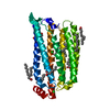
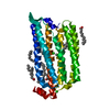
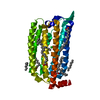
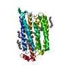
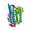
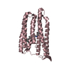
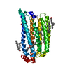
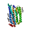
| Title | Structure of Chloride ion pumping rhodopsin (ClR) with NTQ motif 100 ps after light activation (2.63mJ/mm2) | ||||||||||||
 Components Components | Chloride pumping rhodopsin | ||||||||||||
 Keywords Keywords | MEMBRANE PROTEIN / chloride ion pumping rhodopsin / SFX / XFEL / ClR / NTQ | ||||||||||||
| Function / homology | Bacteriorhodopsin-like protein / Archaeal/bacterial/fungal rhodopsins / Bacteriorhodopsin-like protein / photoreceptor activity / phototransduction / membrane / OLEIC ACID / RETINAL / Chloride pumping rhodopsin Function and homology information Function and homology information | ||||||||||||
| Biological species |  Nonlabens marinus S1-08 (bacteria) Nonlabens marinus S1-08 (bacteria) | ||||||||||||
| Method |  X-RAY DIFFRACTION / FREE ELECTRON LASER / MOLECULAR REPLACEMENT / Resolution: 1.85 Å X-RAY DIFFRACTION / FREE ELECTRON LASER / MOLECULAR REPLACEMENT / Resolution: 1.85 Å | ||||||||||||
 Authors Authors | Yun, J.H. / Liu, H. / Lee, W.T. / Schmidt, M. | ||||||||||||
| Funding support |  China, China,  Korea, Republic Of, Korea, Republic Of,  United States, 3items United States, 3items
| ||||||||||||
 Citation Citation | Journal: Proc.Natl.Acad.Sci.USA / Year: 2021 Title: Early-stage dynamics of chloride ion-pumping rhodopsin revealed by a femtosecond X-ray laser. Authors: Yun, J.H. / Li, X. / Yue, J. / Park, J.H. / Jin, Z. / Li, C. / Hu, H. / Shi, Y. / Pandey, S. / Carbajo, S. / Boutet, S. / Hunter, M.S. / Liang, M. / Sierra, R.G. / Lane, T.J. / Zhou, L. / ...Authors: Yun, J.H. / Li, X. / Yue, J. / Park, J.H. / Jin, Z. / Li, C. / Hu, H. / Shi, Y. / Pandey, S. / Carbajo, S. / Boutet, S. / Hunter, M.S. / Liang, M. / Sierra, R.G. / Lane, T.J. / Zhou, L. / Weierstall, U. / Zatsepin, N.A. / Ohki, M. / Tame, J.R.H. / Park, S.Y. / Spence, J.C.H. / Zhang, W. / Schmidt, M. / Lee, W. / Liu, H. | ||||||||||||
| History |
|
- Structure visualization
| Structure viewer | Molecule: MolmilJmol/JSmol |
|---|
- Downloads & links
-Download
| PDBx/mmCIF format | 7crx.cif.gz | 86.7 KB | Display | PDBx/mmCIF format |
|---|---|---|---|---|
| PDB format | pdb7crx.ent.gz | 51.3 KB | Display | PDB format |
| PDBx/mmJSON format | 7crx.json.gz | Tree view | PDBx/mmJSON format | |
| Others |  Other downloads Other downloads |
-Validation report
| Arichive directory | https://data.pdbj.org/pub/pdb/validation_reports/cr/7crxftp://data.pdbj.org/pub/pdb/validation_reports/cr/7crx | HTTPS FTP |
|---|
-Related structure data
| Related structure data |  7criC  7crjC  7crkC  7crlC  7crsC  7crtC  7cryC  5g28S S: Starting model for refinement C: citing same article ( |
|---|---|
| Similar structure data |


-Links
 PDBj
PDBj


- Assembly
| Deposited unit | 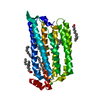
| ||||||||||||
|---|---|---|---|---|---|---|---|---|---|---|---|---|---|
| 1 |
| ||||||||||||
| Unit cell |
|
-Components
| #1: Protein | Mass: 29398.521 Da / Num. of mol.: 1 Source method: isolated from a genetically manipulated source Source: (gene. exp.) Nonlabens marinus S1-08 (bacteria) / Gene: ClR, NMS_1267 / Production host: | ||||||||||
|---|---|---|---|---|---|---|---|---|---|---|---|
| #2: Chemical |   Mass: 35.453 Da / Num. of mol.: 2 / Source method: obtained synthetically / Formula: Cl Mass: 35.453 Da / Num. of mol.: 2 / Source method: obtained synthetically / Formula: Cl#3: Chemical | ChemComp-RET / |   Mass: 284.436 Da / Num. of mol.: 1 / Source method: obtained synthetically / Formula: C20H28O / Feature type: SUBJECT OF INVESTIGATION Mass: 284.436 Da / Num. of mol.: 1 / Source method: obtained synthetically / Formula: C20H28O / Feature type: SUBJECT OF INVESTIGATION#4: Chemical | ChemComp-OLA /   Mass: 282.461 Da / Num. of mol.: 7 / Source method: isolated from a natural source / Formula: C18H34O2 Mass: 282.461 Da / Num. of mol.: 7 / Source method: isolated from a natural source / Formula: C18H34O2#5: Water | ChemComp-HOH / |  Mass: 18.015 Da / Num. of mol.: 106 / Source method: isolated from a natural source / Formula: H2O Mass: 18.015 Da / Num. of mol.: 106 / Source method: isolated from a natural source / Formula: H2OHas ligand of interest | Y | Has protein modification | Y | |
-Experimental details
-Experiment
| Experiment | Method: X-RAY DIFFRACTION / Number of used crystals: 1 |
|---|
- Sample preparation
| Crystal | Density Matthews: 2.85 Å3/Da / Density % sol: 56.85 % |
|---|---|
| Crystal grow | Temperature: 293 K / Method: lipidic cubic phase / pH: 7 / Details: monoolein mixture in syringe |
-Data collection
| Diffraction | Mean temperature: 293 K / Serial crystal experiment: Y |
|---|---|
| Diffraction source | Source: FREE ELECTRON LASER / Site: SLAC LCLS / Beamline: CXI / Wavelength: 1.3 Å |
| Detector | Type: CS-PAD CXI-1 / Detector: PIXEL / Date: Oct 23, 2018 |
| Radiation | Protocol: SINGLE WAVELENGTH / Monochromatic (M) / Laue (L): M / Scattering type: x-ray |
| Radiation wavelength | Wavelength: 1.3 Å / Relative weight: 1 |
| Reflection | Resolution: 1.77→20.9 Å / Num. obs: 33081 / % possible obs: 100 % / Redundancy: 268 % / Biso Wilson estimate: 5.5 Å2 / CC1/2: 0.9786 / CC star: 0.9945 / Net I/σ(I): 5.17 |
| Reflection shell | Resolution: 1.77→1.8 Å / Redundancy: 63.2 % / Mean I/σ(I) obs: 1.54 / Num. unique obs: 1709 / CC1/2: 0.6918 / CC star: 0.9043 / % possible all: 100 |
| Serial crystallography measurement | Pulse duration: 100 fsec. / Pulse photon energy: 9.54 keV |
| Serial crystallography sample delivery | Method: injection |
| Serial crystallography sample delivery injection | Description: LCP injector / Injector diameter: 75 µm |
| Serial crystallography data reduction | Crystal hits: 22474 / Frame hits: 38995 / Frames failed index: 22474 |
- Processing
| Software |
| |||||||||||||||||||||||||||||||||||||||||||||||||||||||||||||||||||||||||||||
|---|---|---|---|---|---|---|---|---|---|---|---|---|---|---|---|---|---|---|---|---|---|---|---|---|---|---|---|---|---|---|---|---|---|---|---|---|---|---|---|---|---|---|---|---|---|---|---|---|---|---|---|---|---|---|---|---|---|---|---|---|---|---|---|---|---|---|---|---|---|---|---|---|---|---|---|---|---|---|
| Refinement | Method to determine structure: MOLECULAR REPLACEMENT Starting model: 5g28 Resolution: 1.85→20.64 Å / SU ML: 0.2901 / Cross valid method: FREE R-VALUE / σ(F): 0 / Phase error: 33.0955 Stereochemistry target values: GeoStd + Monomer Library + CDL v1.2
| |||||||||||||||||||||||||||||||||||||||||||||||||||||||||||||||||||||||||||||
| Solvent computation | Shrinkage radii: 0.9 Å / VDW probe radii: 1.11 Å / Solvent model: FLAT BULK SOLVENT MODEL | |||||||||||||||||||||||||||||||||||||||||||||||||||||||||||||||||||||||||||||
| Displacement parameters | Biso mean: 10.45 Å2 | |||||||||||||||||||||||||||||||||||||||||||||||||||||||||||||||||||||||||||||
| Refinement step | Cycle: LAST / Resolution: 1.85→20.64 Å
| |||||||||||||||||||||||||||||||||||||||||||||||||||||||||||||||||||||||||||||
| Refine LS restraints |
| |||||||||||||||||||||||||||||||||||||||||||||||||||||||||||||||||||||||||||||
| LS refinement shell |
|
