 Movie
Movie Controller


+ Open data

- Basic information
| Entry | Database: PDB / ID: 7cr6 | ||||||
|---|---|---|---|---|---|---|---|
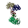
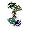
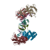
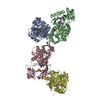
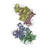
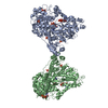
| Title | Synechocystis Cas1-Cas2/prespacer binary complex | ||||||
 Components Components |
| ||||||
 Keywords Keywords | IMMUNE SYSTEM/DNA / CRISPR / adaptation / IMMUNE SYSTEM / IMMUNE SYSTEM-DNA complex | ||||||
| Function / homology |  Function and homology information Function and homology informationCRISPR-cas system / maintenance of CRISPR repeat elements / RNA endonuclease activity / DNA endonuclease activity / endonuclease activity / defense response to virus / Hydrolases; Acting on ester bonds / hydrolase activity / DNA binding / metal ion binding Similarity search - Function | ||||||
| Biological species |  synthetic construct (others) | ||||||
| Method |  X-RAY DIFFRACTION / SYNCHROTRON / MOLECULAR REPLACEMENT / Resolution: 3.72 Å X-RAY DIFFRACTION / SYNCHROTRON / MOLECULAR REPLACEMENT / Resolution: 3.72 Å | ||||||
 Authors Authors | Yu, Y. / Chen, Q. | ||||||
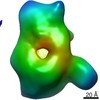
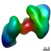
 Citation Citation | Journal: Nucleic Acids Res / Year: 2021 Title: Mechanisms of spacer acquisition by sequential assembly of the adaptation module in Synechocystis. Authors: Chengyong Wu / Dongmei Tang / Jie Cheng / Daojun Hu / Zejing Yang / Xue Ma / Haihuai He / Shaohua Yao / Tian-Min Fu / Yamei Yu / Qiang Chen /   Abstract: CRISPR-Cas immune systems process and integrate short fragments of DNA from new invaders as spacers into the host CRISPR locus to establish molecular memory of prior infection, which is also known as ...CRISPR-Cas immune systems process and integrate short fragments of DNA from new invaders as spacers into the host CRISPR locus to establish molecular memory of prior infection, which is also known as adaptation in the field. Some CRISPR-Cas systems rely on Cas1 and Cas2 to complete the adaptation process, which has been characterized in a few systems. In contrast, many other CRISPR-Cas systems require an additional factor of Cas4 for efficient adaptation, the mechanism of which remains less understood. Here we present biochemical reconstitution of the Synechocystis sp. PCC6803 type I-D adaptation system, X-ray crystal structures of Cas1-Cas2-prespacer complexes, and negative stained electron microscopy structure of the Cas4-Cas1 complex. Cas4 and Cas2 compete with each other to interact with Cas1. In the absence of prespacer, Cas4 but not Cas2 assembles with Cas1 into a very stable complex for processing the prespacer. Strikingly, the Cas1-prespacer complex develops a higher binding affinity toward Cas2 to form the Cas1-Cas2-prespacer ternary complex for integration. Together, we show a two-step sequential assembly mechanism for the type I-D adaptation module of Synechocystis, in which Cas4-Cas1 and Cas1-Cas2 function as two exclusive complexes for prespacer processing, capture, and integration. | ||||||
| History |
|
- Structure visualization
| Structure viewer | Molecule: MolmilJmol/JSmol |
|---|
- Downloads & links
-Download
| PDBx/mmCIF format | 7cr6.cif.gz | 697.3 KB | Display | PDBx/mmCIF format |
|---|---|---|---|---|
| PDB format | pdb7cr6.ent.gz | 468.5 KB | Display | PDB format |
| PDBx/mmJSON format | 7cr6.json.gz | Tree view | PDBx/mmJSON format | |
| Others |  Other downloads Other downloads |
-Validation report
| Arichive directory | https://data.pdbj.org/pub/pdb/validation_reports/cr/7cr6ftp://data.pdbj.org/pub/pdb/validation_reports/cr/7cr6 | HTTPS FTP |
|---|
-Related structure data
| Related structure data | 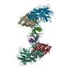 7cr8C  1zpwS S: Starting model for refinement C: citing same article ( |
|---|---|
| Similar structure data |

-Links
 PDBj
PDBj


- Assembly
| Deposited unit | 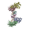
| ||||||||||
|---|---|---|---|---|---|---|---|---|---|---|---|
| 1 |
| ||||||||||
| Unit cell |
|
-Components
| #1: Protein | Mass: 37811.238 Da / Num. of mol.: 4 Source method: isolated from a genetically manipulated source Source: (gene. exp.) Strain: PCC 6803 / Kazusa / Gene: cas1, slr7016 / Production host: References: UniProt: Q6ZEI2, Hydrolases; Acting on ester bonds #2: Protein | Mass: 11656.425 Da / Num. of mol.: 2 Source method: isolated from a genetically manipulated source Source: (gene. exp.) Strain: PCC 6803 / Kazusa / Gene: cas2-1, ssr7017 / Production host: References: UniProt: Q6ZEI1, Hydrolases; Acting on ester bonds #3: DNA chain | | Mass: 10960.986 Da / Num. of mol.: 1 / Source method: obtained synthetically / Source: (synth.) synthetic construct (others) #4: DNA chain | | Mass: 11064.108 Da / Num. of mol.: 1 / Source method: obtained synthetically / Source: (synth.) synthetic construct (others) #5: Water | ChemComp-HOH / |  Mass: 18.015 Da / Num. of mol.: 1 / Source method: isolated from a natural source / Formula: H2O Mass: 18.015 Da / Num. of mol.: 1 / Source method: isolated from a natural source / Formula: H2O |
|---|
-Experimental details
-Experiment
| Experiment | Method: X-RAY DIFFRACTION / Number of used crystals: 1 |
|---|
- Sample preparation
| Crystal | Density Matthews: 3.32 Å3/Da / Density % sol: 62.99 % |
|---|---|
| Crystal grow | Temperature: 293 K / Method: vapor diffusion, hanging drop Details: 30% MPD, 0.1 M imidazole pH 6.5, 0.2 M (NH4)2SO4, and 10% (w/v) PEG3350 |
-Data collection
| Diffraction | Mean temperature: 100 K / Serial crystal experiment: N |
|---|---|
| Diffraction source | Source: SYNCHROTRON / Site: SSRF / Beamline: BL19U1 / Wavelength: 0.9793 Å |
| Detector | Type: DECTRIS PILATUS 6M / Detector: PIXEL / Date: May 10, 2018 |
| Radiation | Protocol: SINGLE WAVELENGTH / Monochromatic (M) / Laue (L): M / Scattering type: x-ray |
| Radiation wavelength | Wavelength: 0.9793 Å / Relative weight: 1 |
| Reflection | Resolution: 3.7→50 Å / Num. obs: 27252 / % possible obs: 99.4 % / Redundancy: 12.5 % / Biso Wilson estimate: 44.65 Å2 / CC1/2: 0.983 / Net I/σ(I): 11 |
| Reflection shell | Resolution: 3.7→3.79 Å / Num. unique obs: 1782 / CC1/2: 0.521 |
- Processing
| Software |
| |||||||||||||||||||||||||||||||||||||||||||||||||||||||||||||||
|---|---|---|---|---|---|---|---|---|---|---|---|---|---|---|---|---|---|---|---|---|---|---|---|---|---|---|---|---|---|---|---|---|---|---|---|---|---|---|---|---|---|---|---|---|---|---|---|---|---|---|---|---|---|---|---|---|---|---|---|---|---|---|---|---|
| Refinement | Method to determine structure: MOLECULAR REPLACEMENT Starting model: 1ZPW Resolution: 3.72→33.81 Å / SU ML: 0.4946 / Cross valid method: THROUGHOUT / σ(F): 1.34 / Phase error: 33.0009 Stereochemistry target values: GeoStd + Monomer Library + CDL v1.2
| |||||||||||||||||||||||||||||||||||||||||||||||||||||||||||||||
| Solvent computation | Shrinkage radii: 0.9 Å / VDW probe radii: 1.11 Å / Solvent model: FLAT BULK SOLVENT MODEL | |||||||||||||||||||||||||||||||||||||||||||||||||||||||||||||||
| Displacement parameters | Biso mean: 43.12 Å2 | |||||||||||||||||||||||||||||||||||||||||||||||||||||||||||||||
| Refinement step | Cycle: LAST / Resolution: 3.72→33.81 Å
| |||||||||||||||||||||||||||||||||||||||||||||||||||||||||||||||
| Refine LS restraints |
| |||||||||||||||||||||||||||||||||||||||||||||||||||||||||||||||
| LS refinement shell |
|
