 Movie
Movie Controller

[English] 日本語
 Yorodumi
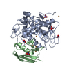
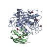
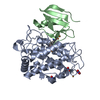
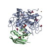
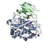
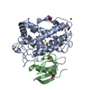
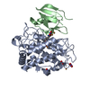
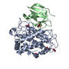
- PDB-7cit: Crystal structure of tyrosinase from Streptomyces castaneoglobisp... -
+ Open data

- Basic information
| Entry | Database: PDB / ID: 7cit | ||||||
|---|---|---|---|---|---|---|---|
| Title | Crystal structure of tyrosinase from Streptomyces castaneoglobisporus in complex with the caddie protein obtained by soaking in the solution containing Cu(II) and hydroxylamine for 24 h | ||||||
 Components Components |
| ||||||
 Keywords Keywords | METAL BINDING PROTEIN / tyrosinase / catalytic mechanism | ||||||
| Function / homology | COPPER (II) ION / NITRATE ION / HYDROGEN PEROXIDE Function and homology information Function and homology information | ||||||
| Biological species |  Streptomyces castaneoglobisporus (bacteria) Streptomyces castaneoglobisporus (bacteria) | ||||||
| Method |  X-RAY DIFFRACTION / SYNCHROTRON / MOLECULAR REPLACEMENT / Resolution: 1.5 Å X-RAY DIFFRACTION / SYNCHROTRON / MOLECULAR REPLACEMENT / Resolution: 1.5 Å | ||||||
| Model details | Tyr98 residue of caddie is partially oxygenated | ||||||
 Authors Authors | Oda, K. / Matoba, Y. | ||||||
 Citation Citation | Journal: Int.J.Biol.Macromol. / Year: 2021 Title: The basicity of an active-site water molecule discriminates between tyrosinase and catechol oxidase activity. Authors: Matoba, Y. / Oda, K. / Muraki, Y. / Masuda, T. | ||||||
| History |
|
- Structure visualization
| Structure viewer | Molecule: MolmilJmol/JSmol |
|---|
- Downloads & links
-Download
| PDBx/mmCIF format | 7cit.cif.gz | 175.1 KB | Display | PDBx/mmCIF format |
|---|---|---|---|---|
| PDB format | pdb7cit.ent.gz | 136 KB | Display | PDB format |
| PDBx/mmJSON format | 7cit.json.gz | Tree view | PDBx/mmJSON format | |
| Others |  Other downloads Other downloads |
-Validation report
| Arichive directory | https://data.pdbj.org/pub/pdb/validation_reports/ci/7citftp://data.pdbj.org/pub/pdb/validation_reports/ci/7cit | HTTPS FTP |
|---|
-Related structure data
| Related structure data |  7ciyC  2zmyS S: Starting model for refinement C: citing same article ( |
|---|---|
| Similar structure data |


-Links
 PDBj
PDBj- Assembly
| Deposited unit | 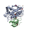
| ||||||||||||
|---|---|---|---|---|---|---|---|---|---|---|---|---|---|
| 1 |
| ||||||||||||
| Unit cell |
| ||||||||||||
| Components on special symmetry positions |
|
-Components
-Protein , 2 types, 2 molecules AB
| #1: Protein | Mass: 32089.564 Da / Num. of mol.: 1 Source method: isolated from a genetically manipulated source Source: (gene. exp.) Streptomyces castaneoglobisporus (bacteria)Strain: HUT 6202 / Plasmid: PET-MEL2 / Production host: |
|---|---|
| #2: Protein | Mass: 14217.795 Da / Num. of mol.: 1 Source method: isolated from a genetically manipulated source Source: (gene. exp.) Streptomyces castaneoglobisporus (bacteria)Strain: HUT 6202 / Plasmid: PET-MEL2 / Production host: |
-Non-polymers , 4 types, 335 molecules 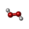


| #3: Chemical | ChemComp-PEO /  Mass: 34.015 Da / Num. of mol.: 1 / Source method: obtained synthetically / Formula: H2O2 / Feature type: SUBJECT OF INVESTIGATION Mass: 34.015 Da / Num. of mol.: 1 / Source method: obtained synthetically / Formula: H2O2 / Feature type: SUBJECT OF INVESTIGATION | ||||
|---|---|---|---|---|---|
| #4: Chemical | ChemComp-CU /  Mass: 63.546 Da / Num. of mol.: 5 / Source method: obtained synthetically / Formula: Cu / Feature type: SUBJECT OF INVESTIGATION Mass: 63.546 Da / Num. of mol.: 5 / Source method: obtained synthetically / Formula: Cu / Feature type: SUBJECT OF INVESTIGATION#5: Chemical | ChemComp-NO3 /  Mass: 62.005 Da / Num. of mol.: 6 / Source method: obtained synthetically / Formula: NO3 Mass: 62.005 Da / Num. of mol.: 6 / Source method: obtained synthetically / Formula: NO3#6: Water | ChemComp-HOH / | Mass: 18.015 Da / Num. of mol.: 323 / Source method: isolated from a natural source / Formula: H2O |
-Details
| Has ligand of interest | Y |
|---|
-Experimental details
-Experiment
| Experiment | Method: X-RAY DIFFRACTION / Number of used crystals: 1 |
|---|
- Sample preparation
| Crystal | Density Matthews: 1.84 Å3/Da / Density % sol: 32.61 % / Mosaicity: 0.824 ° |
|---|---|
| Crystal grow | Temperature: 298 K / Method: vapor diffusion, sitting drop / pH: 6.5 / Details: PEG 3350, NaNO3 |
-Data collection
| Diffraction | Mean temperature: 100 K / Serial crystal experiment: N |
|---|---|
| Diffraction source | Source: SYNCHROTRON / Site: SPring-8  / Beamline: BL26B2 / Wavelength: 0.9 Å / Beamline: BL26B2 / Wavelength: 0.9 Å |
| Detector | Type: RAYONIX MX-225 / Detector: CCD / Date: Nov 17, 2010 |
| Radiation | Protocol: SINGLE WAVELENGTH / Monochromatic (M) / Laue (L): M / Scattering type: x-ray |
| Radiation wavelength | Wavelength: 0.9 Å / Relative weight: 1 |
| Reflection | Resolution: 1.5→100 Å / Num. obs: 53168 / % possible obs: 96.3 % / Redundancy: 6.3 % / Rmerge(I) obs: 0.049 / Χ2: 0.982 / Net I/σ(I): 35.6 |
| Reflection shell | Resolution: 1.5→1.55 Å / Rmerge(I) obs: 0.432 / Num. unique obs: 5762 / Χ2: 1.176 |
- Processing
| Software |
| |||||||||||||||||||||||||||||||||
|---|---|---|---|---|---|---|---|---|---|---|---|---|---|---|---|---|---|---|---|---|---|---|---|---|---|---|---|---|---|---|---|---|---|---|
| Refinement | Method to determine structure: MOLECULAR REPLACEMENT Starting model: 2ZMY Resolution: 1.5→30 Å / Cross valid method: FREE R-VALUE / σ(F): 0 / Stereochemistry target values: ENGH AND HUBER Details: ANISOTROPIC REFINEMENT REDUCED FREE R (NO CUTOFF) BY ?
| |||||||||||||||||||||||||||||||||
| Solvent computation | Solvent model: MOEWS & KRETSINGER, J.MOL.BIOL.91(1973)201-228 | |||||||||||||||||||||||||||||||||
| Displacement parameters | Biso max: 129.37 Å2 / Biso mean: 25.7909 Å2 / Biso min: 12.19 Å2 | |||||||||||||||||||||||||||||||||
| Refinement step | Cycle: final / Resolution: 1.5→30 Å
| |||||||||||||||||||||||||||||||||
| Refine LS restraints |
|
