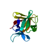
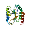
- PDB-7c1j: Crystal structure of the receiver domain of sensor histidine kina... -
+
Open data
ID or keywords:
Loading...
-
Basic information
Entry
Database: PDB / ID: 7c1j
Title
Crystal structure of the receiver domain of sensor histidine kinase PA1611 (PA1611REC) from Pseudomonas aeruginosa PAO1 with magnesium ion coordinated in the active site cleft
Components
Histidine kinase
Keywords
TRANSFERASE / P. aeruginosa / Two-component regulatory system / Sensor histidine kinase / Histidine-containing phosphotransfer protein
Function / homology
Function and homology information
histidine phosphotransfer kinase activity / phosphorelay sensor kinase activity / histidine kinase / phosphorelay signal transduction system / ATP binding / metal ion binding / plasma membrane Similarity search - Function
Resolution: 1.35→20.16 Å / Cor.coef. Fo:Fc: 0.967 / Cor.coef. Fo:Fc free: 0.953 / SU B: 1.244 / SU ML: 0.049 / Cross valid method: THROUGHOUT / ESU R: 0.061 / ESU R Free: 0.066 / Stereochemistry target values: MAXIMUM LIKELIHOOD / Details: HYDROGENS HAVE BEEN ADDED IN THE RIDING POSITIONS
Rfactor
Num. reflection
% reflection
Selection details
Rfree
0.21637
1448
5.1 %
RANDOM
Rwork
0.18038
-
-
-
obs
0.18217
26942
96.99 %
-
Solvent computation
Ion probe radii: 0.8 Å / Shrinkage radii: 0.8 Å / VDW probe radii: 1.2 Å / Solvent model: MASK
 Movie
Movie Controller

 Yorodumi
 Open data
 Basic information
 Components
 Keywords
 Function and homology information
 Pseudomonas aeruginosa (bacteria)
 X-RAY DIFFRACTION /
  Authors
 Citation
 Structure visualization
 Downloads & links
 Other downloads
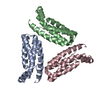


 PDBj
PDBj


 Assembly
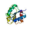

 Mass: 24.305 Da / Num. of mol.: 1 / Source method: obtained synthetically / Formula: Mg / Feature type: SUBJECT OF INVESTIGATION
 Mass: 18.015 Da / Num. of mol.: 173 / Source method: isolated from a natural source / Formula: H2O
 Sample preparation
 / Beamline: BL15A1 / Wavelength: 1 Å
 Processing