 Movie
Movie Controller

[English] 日本語
 Yorodumi
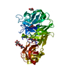
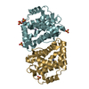
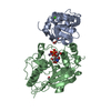
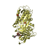
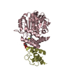
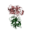
- PDB-7byt: Crystal structure of exo-beta-1,3-galactanase from Phanerochaete ... -
+ Open data

- Basic information
| Entry | Database: PDB / ID: 7byt | ||||||
|---|---|---|---|---|---|---|---|
| Title | Crystal structure of exo-beta-1,3-galactanase from Phanerochaete chrysosporium Pc1,3Gal43A with galactose | ||||||
 Components Components | Galactan 1,3-beta-galactosidase | ||||||
 Keywords Keywords | HYDROLASE / Glycoside hydrolase family 43 / exo-beta-1 / 3-galactanase / arabinogalactan degrade / complex with galactotriose / carbohydrate binding module family 35 | ||||||
| Function / homology |  Function and homology information Function and homology informationgalactan 1,3-beta-galactosidase / galactan 1,3-beta-galactosidase activity / carbohydrate binding / carbohydrate metabolic process / metal ion binding Similarity search - Function | ||||||
| Biological species |  Phanerochaete chrysosporium (fungus) Phanerochaete chrysosporium (fungus) | ||||||
| Method |  X-RAY DIFFRACTION / SYNCHROTRON / MOLECULAR REPLACEMENT / Resolution: 1.5 Å X-RAY DIFFRACTION / SYNCHROTRON / MOLECULAR REPLACEMENT / Resolution: 1.5 Å | ||||||
 Authors Authors | Matsuyama, K. / Ishida, T. / Kishine, N. / Fujimoto, Z. / Igarashi, K. / Kaneko, S. | ||||||
| Funding support |  Japan, 1items Japan, 1items
| ||||||
 Citation Citation | Journal: J.Biol.Chem. / Year: 2020 Title: Unique active-site and subsite features in the arabinogalactan-degrading GH43 exo-beta-1,3-galactanase from Phanerochaete chrysosporium . Authors: Matsuyama, K. / Kishine, N. / Fujimoto, Z. / Sunagawa, N. / Kotake, T. / Tsumuraya, Y. / Samejima, M. / Igarashi, K. / Kaneko, S. | ||||||
| History |
|
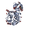
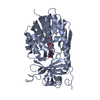
- Structure visualization
| Structure viewer | Molecule: MolmilJmol/JSmol |
|---|
- Downloads & links
-Download
| PDBx/mmCIF format | 7byt.cif.gz | 116.4 KB | Display | PDBx/mmCIF format |
|---|---|---|---|---|
| PDB format | pdb7byt.ent.gz | 83.1 KB | Display | PDB format |
| PDBx/mmJSON format | 7byt.json.gz | Tree view | PDBx/mmJSON format | |
| Others |  Other downloads Other downloads |
-Validation report
| Arichive directory | https://data.pdbj.org/pub/pdb/validation_reports/by/7bytftp://data.pdbj.org/pub/pdb/validation_reports/by/7byt | HTTPS FTP |
|---|
-Related structure data


-Links
 PDBj
PDBj


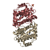
- Assembly
| Deposited unit | 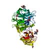
| ||||||||||
|---|---|---|---|---|---|---|---|---|---|---|---|
| 1 |
| ||||||||||
| Unit cell |
|
-Components
-Protein , 1 types, 1 molecules A
| #1: Protein | Mass: 45535.648 Da / Num. of mol.: 1 Source method: isolated from a genetically manipulated source Source: (gene. exp.) Phanerochaete chrysosporium (fungus) / Strain: K-3 / Plasmid: pPICZalphaA / Production host: Komagataella pastoris (fungus) / Strain (production host): KM71HReferences: UniProt: Q50KB2, galactan 1,3-beta-galactosidase |
|---|
-Sugars , 4 types, 5 molecules 
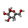

| #2: Polysaccharide | 2-acetamido-2-deoxy-beta-D-glucopyranose-(1-4)-2-acetamido-2-deoxy-beta-D-glucopyranose Source method: isolated from a genetically manipulated source | ||||
|---|---|---|---|---|---|
| #3: Sugar |  Type: D-saccharide, beta linking / Mass: 221.208 Da / Num. of mol.: 2 Type: D-saccharide, beta linking / Mass: 221.208 Da / Num. of mol.: 2Source method: isolated from a genetically manipulated source Formula: C8H15NO6 #5: Sugar | ChemComp-GLA / |  Type: D-saccharide, alpha linking / Mass: 180.156 Da / Num. of mol.: 1 Type: D-saccharide, alpha linking / Mass: 180.156 Da / Num. of mol.: 1Source method: isolated from a genetically manipulated source Formula: C6H12O6 / Feature type: SUBJECT OF INVESTIGATION #6: Sugar | ChemComp-GAL / |  Type: D-saccharide, beta linking / Mass: 180.156 Da / Num. of mol.: 1 Type: D-saccharide, beta linking / Mass: 180.156 Da / Num. of mol.: 1Source method: isolated from a genetically manipulated source Formula: C6H12O6 / Feature type: SUBJECT OF INVESTIGATION |
-Non-polymers , 5 types, 521 molecules 


| #4: Chemical | ChemComp-CA /  Mass: 40.078 Da / Num. of mol.: 1 Mass: 40.078 Da / Num. of mol.: 1Source method: isolated from a genetically manipulated source Formula: Ca | ||||||
|---|---|---|---|---|---|---|---|
| #7: Chemical |  Mass: 60.052 Da / Num. of mol.: 2 / Source method: obtained synthetically / Formula: C2H4O2 Mass: 60.052 Da / Num. of mol.: 2 / Source method: obtained synthetically / Formula: C2H4O2#8: Chemical |  Mass: 59.044 Da / Num. of mol.: 2 / Source method: isolated from a natural source / Formula: C2H3O2 Mass: 59.044 Da / Num. of mol.: 2 / Source method: isolated from a natural source / Formula: C2H3O2#9: Chemical | ChemComp-GOL /  Mass: 92.094 Da / Num. of mol.: 4 / Source method: isolated from a natural source / Formula: C3H8O3 Mass: 92.094 Da / Num. of mol.: 4 / Source method: isolated from a natural source / Formula: C3H8O3#10: Water | ChemComp-HOH / | Mass: 18.015 Da / Num. of mol.: 512 / Source method: isolated from a natural source / Formula: H2O |
-Details
| Has ligand of interest | Y |
|---|---|
| Has protein modification | Y |
-Experimental details
-Experiment
| Experiment | Method: X-RAY DIFFRACTION / Number of used crystals: 1 |
|---|
- Sample preparation
| Crystal | Density Matthews: 1.98 Å3/Da / Density % sol: 37.75 % |
|---|---|
| Crystal grow | Temperature: 293 K / Method: vapor diffusion, hanging drop / pH: 5.5 Details: 16 % (w/v) polyethylene glycol (PEG) 10000, 0.1 M ammonium sulfate, 0.1 M bis-tris (pH 5.5), 5 % (v/v) glycerol, Crystal was soaked into 1 % (w/v) beta-1,3-galactotriose for 5 min |
-Data collection
| Diffraction | Mean temperature: 95 K / Serial crystal experiment: N |
|---|---|
| Diffraction source | Source: SYNCHROTRON / Site: Photon Factory / Beamline: AR-NW12A / Wavelength: 1 Å |
| Detector | Type: ADSC QUANTUM 210 / Detector: CCD / Date: Dec 7, 2008 |
| Radiation | Monochromator: 1 / Protocol: SINGLE WAVELENGTH / Monochromatic (M) / Laue (L): M / Scattering type: x-ray |
| Radiation wavelength | Wavelength: 1 Å / Relative weight: 1 |
| Reflection | Resolution: 1.5→100 Å / Num. obs: 57278 / % possible obs: 97.5 % / Redundancy: 9.2 % / Biso Wilson estimate: 10.11 Å2 / Rsym value: 0.046 / Net I/σ(I): 48.9 |
| Reflection shell | Resolution: 1.5→1.55 Å / Redundancy: 8.9 % / Mean I/σ(I) obs: 21 / Num. unique obs: 5493 / Rsym value: 0.109 / % possible all: 94.9 |
- Processing
| Software |
| |||||||||||||||||||||
|---|---|---|---|---|---|---|---|---|---|---|---|---|---|---|---|---|---|---|---|---|---|---|
| Refinement | Method to determine structure: MOLECULAR REPLACEMENT Starting model: D_1300016321 Resolution: 1.5→41.56 Å / Cross valid method: FREE R-VALUE
| |||||||||||||||||||||
| Displacement parameters | Biso max: 55.9 Å2 / Biso mean: 12.4109 Å2 / Biso min: 4.96 Å2 | |||||||||||||||||||||
| Refinement step | Cycle: LAST / Resolution: 1.5→41.56 Å
| |||||||||||||||||||||
| LS refinement shell | Resolution: 1.5→1.554 Å /
|
