 Movie
Movie Controller

[English] 日本語
 Yorodumi
- PDB-7bvt: Crystal structure of cyclic alpha-maltosyl-1,6-maltose binding pr... -
+ Open data

- Basic information
| Entry | Database: PDB / ID: 7bvt | ||||||
|---|---|---|---|---|---|---|---|
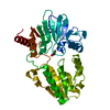
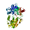
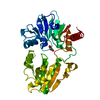
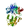
| Title | Crystal structure of cyclic alpha-maltosyl-1,6-maltose binding protein from Arthrobacter globiformis | ||||||
 Components Components | Hypothetical sugar ABC-transporter sugar binding protein | ||||||
 Keywords Keywords | SUGAR BINDING PROTEIN / substrate-binding protein / ABC transporter | ||||||
| Function / homology | : / Bacterial extracellular solute-binding protein / Solute-binding family 1, conserved site / Bacterial extracellular solute-binding proteins, family 1 signature. / Bacterial extracellular solute-binding protein / transmembrane transport / Prokaryotic membrane lipoprotein lipid attachment site profile. / Hypothetical sugar ABC-transporter sugar binding protein Function and homology information Function and homology information | ||||||
| Biological species |  Arthrobacter globiformis (bacteria) Arthrobacter globiformis (bacteria) | ||||||
| Method |  X-RAY DIFFRACTION / SYNCHROTRON / MOLECULAR REPLACEMENT / Resolution: 1.47 Å X-RAY DIFFRACTION / SYNCHROTRON / MOLECULAR REPLACEMENT / Resolution: 1.47 Å | ||||||
 Authors Authors | Kohno, M. / Arakawa, T. / Mori, T. / Nishimoto, T. / Fushinobu, S. | ||||||
| Funding support |  Japan, 1items Japan, 1items
| ||||||
 Citation Citation | Journal: Plos One / Year: 2020 Title: Molecular analysis of cyclic alpha-maltosyl-(1→6)-maltose binding protein in the bacterial metabolic pathway. Authors: Kohno, M. / Arakawa, T. / Sunagawa, N. / Mori, T. / Igarashi, K. / Nishimoto, T. / Fushinobu, S. #1: Journal: Journal of Applied Glycoscience / Year: 2011Title: Cloning, Sequencing and Expression of the Genes Encoding Cyclic alpha-Maltosyl-(1->6)-maltose Hydrolase and alpha-Glucosidase from an Arthrobacter globiformis Strain Authors: Mori, T. / Nishimoto, T. / Okura, T. / Chaen, H. / Fukuda, S. | ||||||
| History |
|
- Structure visualization
| Structure viewer | Molecule: MolmilJmol/JSmol |
|---|
- Downloads & links
-Download
| PDBx/mmCIF format | 7bvt.cif.gz | 99.4 KB | Display | PDBx/mmCIF format |
|---|---|---|---|---|
| PDB format | pdb7bvt.ent.gz | 72.5 KB | Display | PDB format |
| PDBx/mmJSON format | 7bvt.json.gz | Tree view | PDBx/mmJSON format | |
| Others |  Other downloads Other downloads |
-Validation report
| Arichive directory | https://data.pdbj.org/pub/pdb/validation_reports/bv/7bvtftp://data.pdbj.org/pub/pdb/validation_reports/bv/7bvt | HTTPS FTP |
|---|
-Related structure data
| Related structure data |  5ci5S S: Starting model for refinement |
|---|---|
| Similar structure data |


-Links
 PDBj
PDBj


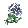
- Assembly
| Deposited unit | 
| ||||||||
|---|---|---|---|---|---|---|---|---|---|
| 1 |
| ||||||||
| Unit cell |
|
-Components
| #1: Protein | Mass: 44495.172 Da / Num. of mol.: 1 / Fragment: substrate binding protein Source method: isolated from a genetically manipulated source Source: (gene. exp.) Arthrobacter globiformis (bacteria) / Gene: cmmC / Plasmid: pET-28b / Production host: |
|---|---|
| #2: Polysaccharide | alpha-D-glucopyranose-(1-6)-alpha-D-glucopyranose-(1-4)-alpha-D-glucopyranose Source method: isolated from a genetically manipulated source |
| #3: Water | ChemComp-HOH /  Mass: 18.015 Da / Num. of mol.: 381 / Source method: isolated from a natural source / Formula: H2O Mass: 18.015 Da / Num. of mol.: 381 / Source method: isolated from a natural source / Formula: H2O |
| Has ligand of interest | Y |
-Experimental details
-Experiment
| Experiment | Method: X-RAY DIFFRACTION / Number of used crystals: 1 |
|---|
- Sample preparation
| Crystal | Density Matthews: 2.11 Å3/Da / Density % sol: 41.69 % |
|---|---|
| Crystal grow | Temperature: 293 K / Method: vapor diffusion, sitting drop / pH: 8.5 / Details: 0.1 M Tris-HCl (pH 8.5), 2.0 M ammonium sulfate |
-Data collection
| Diffraction | Mean temperature: 100 K / Serial crystal experiment: N |
|---|---|
| Diffraction source | Source: SYNCHROTRON / Site: Photon Factory / Beamline: AR-NW12A / Wavelength: 1 Å |
| Detector | Type: DECTRIS PILATUS3 S 2M / Detector: PIXEL / Date: Nov 18, 2017 |
| Radiation | Monochromator: Numerical link type Si(111) double crystal / Protocol: SINGLE WAVELENGTH / Monochromatic (M) / Laue (L): M / Scattering type: x-ray |
| Radiation wavelength | Wavelength: 1 Å / Relative weight: 1 |
| Reflection | Resolution: 1.47→46.04 Å / Num. obs: 68976 / % possible obs: 99.5 % / Redundancy: 19.6 % / CC1/2: 0.981 / Rmerge(I) obs: 0.093 / Rpim(I) all: 0.021 / Net I/σ(I): 15.7 |
| Reflection shell | Resolution: 1.47→1.5 Å / Redundancy: 10 % / Rmerge(I) obs: 1.794 / Mean I/σ(I) obs: 1.2 / Num. unique obs: 32216 / CC1/2: 0.93 / Rpim(I) all: 0.595 / % possible all: 95.8 |
- Processing
| Software |
| ||||||||||||||||||||||||||||||||||||||||||||||||||||||||||||
|---|---|---|---|---|---|---|---|---|---|---|---|---|---|---|---|---|---|---|---|---|---|---|---|---|---|---|---|---|---|---|---|---|---|---|---|---|---|---|---|---|---|---|---|---|---|---|---|---|---|---|---|---|---|---|---|---|---|---|---|---|---|
| Refinement | Method to determine structure: MOLECULAR REPLACEMENT Starting model: 5CI5 Resolution: 1.47→44.67 Å / Cor.coef. Fo:Fc: 0.954 / Cor.coef. Fo:Fc free: 0.963 / SU B: 1.748 / SU ML: 0.061 / Cross valid method: THROUGHOUT / σ(F): 0 / ESU R: 0.07 / ESU R Free: 0.07 / Stereochemistry target values: MAXIMUM LIKELIHOOD Details: HYDROGENS HAVE BEEN ADDED IN THE RIDING POSITIONS U VALUES : REFINED INDIVIDUALLY
| ||||||||||||||||||||||||||||||||||||||||||||||||||||||||||||
| Solvent computation | Ion probe radii: 0.8 Å / Shrinkage radii: 0.8 Å / VDW probe radii: 1.2 Å / Solvent model: BABINET MODEL WITH MASK | ||||||||||||||||||||||||||||||||||||||||||||||||||||||||||||
| Displacement parameters | Biso max: 75.34 Å2 / Biso mean: 22.716 Å2 / Biso min: 8.4 Å2
| ||||||||||||||||||||||||||||||||||||||||||||||||||||||||||||
| Refinement step | Cycle: final / Resolution: 1.47→44.67 Å
| ||||||||||||||||||||||||||||||||||||||||||||||||||||||||||||
| Refine LS restraints |
| ||||||||||||||||||||||||||||||||||||||||||||||||||||||||||||
| LS refinement shell | Resolution: 1.47→1.508 Å / Rfactor Rfree error: 0 / Total num. of bins used: 20
|
