 Movie
Movie Controller


+ Open data

- Basic information
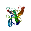
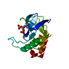
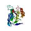
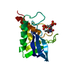
| Entry | Database: PDB / ID: 7bv0 | ||||||
|---|---|---|---|---|---|---|---|
| Title | HEV-C E2s | ||||||
 Components Components | Protein ORF2 | ||||||
 Keywords Keywords | VIRAL PROTEIN / ORF2 / E2S | ||||||
| Function / homology |  Function and homology information Function and homology informationT=1 icosahedral viral capsid / host cell endoplasmic reticulum / host cell surface / host cell Golgi apparatus / structural molecule activity / RNA binding / extracellular region Similarity search - Function | ||||||
| Biological species |  Hepatitis E virus Hepatitis E virus | ||||||
| Method |  X-RAY DIFFRACTION / FREE ELECTRON LASER / MOLECULAR REPLACEMENT / Resolution: 1.801 Å X-RAY DIFFRACTION / FREE ELECTRON LASER / MOLECULAR REPLACEMENT / Resolution: 1.801 Å | ||||||
 Authors Authors | Bai, C.Z. / Qi, J.X. | ||||||
 Citation Citation | Journal: To Be Published Title: Structure of emerging human-infected hepatitis E virus E2s at 1.8 Angstroms resolution Authors: Bai, C.Z. / Qi, J.X. / Wang, Q.H. | ||||||
| History |
|
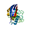
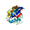
- Structure visualization
| Structure viewer | Molecule: MolmilJmol/JSmol |
|---|
- Downloads & links
-Download
| PDBx/mmCIF format | 7bv0.cif.gz | 85.1 KB | Display | PDBx/mmCIF format |
|---|---|---|---|---|
| PDB format | pdb7bv0.ent.gz | 61.9 KB | Display | PDB format |
| PDBx/mmJSON format | 7bv0.json.gz | Tree view | PDBx/mmJSON format | |
| Others |  Other downloads Other downloads |
-Validation report
| Arichive directory | https://data.pdbj.org/pub/pdb/validation_reports/bv/7bv0ftp://data.pdbj.org/pub/pdb/validation_reports/bv/7bv0 | HTTPS FTP |
|---|
-Related structure data
| Related structure data |  3ggqS S: Starting model for refinement |
|---|---|
| Similar structure data |


-Links
 PDBj
PDBj
- Assembly
| Deposited unit | 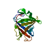
| |||||||||||||||||||||
|---|---|---|---|---|---|---|---|---|---|---|---|---|---|---|---|---|---|---|---|---|---|---|
| 1 |
| |||||||||||||||||||||
| Unit cell |
| |||||||||||||||||||||
| Components on special symmetry positions |
|
-Components
| #1: Protein | Mass: 25772.547 Da / Num. of mol.: 1 Source method: isolated from a genetically manipulated source Source: (gene. exp.) Hepatitis E virus / Production host:  |
|---|---|
| #2: Water | ChemComp-HOH /  Mass: 18.015 Da / Num. of mol.: 237 / Source method: isolated from a natural source / Formula: H2O Mass: 18.015 Da / Num. of mol.: 237 / Source method: isolated from a natural source / Formula: H2O |
-Experimental details
-Experiment
| Experiment | Method: X-RAY DIFFRACTION / Number of used crystals: 1 |
|---|
- Sample preparation
| Crystal | Density Matthews: 2.45 Å3/Da / Density % sol: 49.78 % |
|---|---|
| Crystal grow | Temperature: 293.5 K / Method: batch mode Details: 0.1 M sodium acetate trihydrate pH 4.5, 3.0 M sodium chloride |
-Data collection
| Diffraction | Mean temperature: 293 K / Serial crystal experiment: N |
|---|---|
| Diffraction source | Source: FREE ELECTRON LASER / Site: PAL-XFEL  / Beamline: NCI / Wavelength: 0.988 Å / Beamline: NCI / Wavelength: 0.988 Å |
| Detector | Type: Nonius Kappa CCD / Detector: CCD / Date: Oct 9, 2019 |
| Radiation | Protocol: SINGLE WAVELENGTH / Monochromatic (M) / Laue (L): M / Scattering type: x-ray |
| Radiation wavelength | Wavelength: 0.988 Å / Relative weight: 1 |
| Reflection | Resolution: 1.801→46.618 Å / Num. obs: 24297 / % possible obs: 99.74 % / Redundancy: 20 % / CC1/2: 0.75 / Net I/σ(I): 1 |
| Reflection shell | Resolution: 1.801→1.8736 Å / Num. unique obs: 24297 / CC1/2: 0.5 / % possible all: 99 |
- Processing
| Software |
| ||||||||||||||||||||||||||||||||||||||||||||||||||||||||||||
|---|---|---|---|---|---|---|---|---|---|---|---|---|---|---|---|---|---|---|---|---|---|---|---|---|---|---|---|---|---|---|---|---|---|---|---|---|---|---|---|---|---|---|---|---|---|---|---|---|---|---|---|---|---|---|---|---|---|---|---|---|---|
| Refinement | Method to determine structure: MOLECULAR REPLACEMENT Starting model: 3GGQ Resolution: 1.801→46.618 Å / SU ML: 0.14 / Cross valid method: THROUGHOUT / σ(F): 1.36 / Phase error: 16.73
| ||||||||||||||||||||||||||||||||||||||||||||||||||||||||||||
| Solvent computation | Shrinkage radii: 0.9 Å / VDW probe radii: 1.11 Å | ||||||||||||||||||||||||||||||||||||||||||||||||||||||||||||
| Displacement parameters | Biso max: 81.67 Å2 / Biso mean: 22.7474 Å2 / Biso min: 10.15 Å2 | ||||||||||||||||||||||||||||||||||||||||||||||||||||||||||||
| Refinement step | Cycle: final / Resolution: 1.801→46.618 Å
| ||||||||||||||||||||||||||||||||||||||||||||||||||||||||||||
| LS refinement shell | Refine-ID: X-RAY DIFFRACTION / Rfactor Rfree error: 0
| ||||||||||||||||||||||||||||||||||||||||||||||||||||||||||||
| Refinement TLS params. | Method: refined / Origin x: -11.7706 Å / Origin y: -37.79 Å / Origin z: -9.1327 Å
| ||||||||||||||||||||||||||||||||||||||||||||||||||||||||||||
| Refinement TLS group |
|
