 Movie
Movie Controller

[English] 日本語
 Yorodumi
- PDB-7bui: Complex of reduced oxygenase and oxidized ferredoxin in carbazole... -
+ Open data

- Basic information
| Entry | Database: PDB / ID: 7bui | ||||||
|---|---|---|---|---|---|---|---|
| Title | Complex of reduced oxygenase and oxidized ferredoxin in carbazole 1,9a- dioxygenase | ||||||
 Components Components |
| ||||||
 Keywords Keywords | OXIDOREDUCTASE / Rieske Iron-sulfur Protein | ||||||
| Function / homology |  Function and homology information Function and homology informationcarbazole catabolic process / ferredoxin hydrogenase activity / dioxygenase activity / 2 iron, 2 sulfur cluster binding / oxidoreductase activity / metal ion binding Similarity search - Function | ||||||
| Biological species | Janthinobacterium sp. Pseudomonas resinovorans (bacteria) Pseudomonas resinovorans (bacteria) | ||||||
| Method |  X-RAY DIFFRACTION / SYNCHROTRON / MOLECULAR REPLACEMENT / Resolution: 2.15 Å X-RAY DIFFRACTION / SYNCHROTRON / MOLECULAR REPLACEMENT / Resolution: 2.15 Å | ||||||
 Authors Authors | Matsuzawa, J. / Wang, Y.X. / Suzuki-Minakuchi, C. / Nojiri, H. | ||||||
| Funding support |  Japan, 1items Japan, 1items
| ||||||
 Citation Citation | Journal: To Be Published Title: Complex of reduced oxygenase and oxidized ferredoxin in carbazole 1,9a- dioxygenase Authors: Matsuzawa, J. / Wang, Y.X. / Suzuki-Minakuchi, C. / Nojiri, H. | ||||||
| History |
|
- Structure visualization
| Structure viewer | Molecule: MolmilJmol/JSmol |
|---|
- Downloads & links
-Download
| PDBx/mmCIF format | 7bui.cif.gz | 296.1 KB | Display | PDBx/mmCIF format |
|---|---|---|---|---|
| PDB format | pdb7bui.ent.gz | 236.9 KB | Display | PDB format |
| PDBx/mmJSON format | 7bui.json.gz | Tree view | PDBx/mmJSON format | |
| Others |  Other downloads Other downloads |
-Validation report
| Summary document | 7bui_validation.pdf.gz | 1.7 MB | Display | wwPDB validaton report |
|---|---|---|---|---|
| Full document | 7bui_full_validation.pdf.gz | 1.7 MB | Display | |
| Data in XML | 7bui_validation.xml.gz | 53.8 KB | Display | |
| Data in CIF | 7bui_validation.cif.gz | 76.3 KB | Display | |
| Arichive directory | https://data.pdbj.org/pub/pdb/validation_reports/bu/7buiftp://data.pdbj.org/pub/pdb/validation_reports/bu/7bui | HTTPS FTP |
-Related structure data
| Related structure data |  2de5S S: Starting model for refinement |
|---|---|
| Similar structure data |


-Links
 PDBj
PDBj


- Assembly
| Deposited unit | 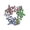
| ||||||||
|---|---|---|---|---|---|---|---|---|---|
| 1 |
| ||||||||
| Unit cell |
|
-Components
-Protein , 2 types, 5 molecules ABCDE
| #1: Protein | Mass: 44910.738 Da / Num. of mol.: 3 Source method: isolated from a genetically manipulated source Source: (gene. exp.) Janthinobacterium sp. (strain J3) (bacteria)Strain: J3 / Gene: carAa / Production host: #2: Protein | Mass: 12448.059 Da / Num. of mol.: 2 Source method: isolated from a genetically manipulated source Source: (gene. exp.) Pseudomonas resinovorans (bacteria) / Gene: carAc / Production host: |
|---|
-Non-polymers , 5 types, 497 molecules 


| #3: Chemical | ChemComp-FES /  Mass: 175.820 Da / Num. of mol.: 5 / Source method: obtained synthetically / Formula: Fe2S2 Mass: 175.820 Da / Num. of mol.: 5 / Source method: obtained synthetically / Formula: Fe2S2#4: Chemical |  Mass: 55.845 Da / Num. of mol.: 3 / Source method: obtained synthetically / Formula: Fe / Feature type: SUBJECT OF INVESTIGATION Mass: 55.845 Da / Num. of mol.: 3 / Source method: obtained synthetically / Formula: Fe / Feature type: SUBJECT OF INVESTIGATION#5: Chemical | ChemComp-EDO /  Mass: 62.068 Da / Num. of mol.: 10 / Source method: obtained synthetically / Formula: C2H6O2 Mass: 62.068 Da / Num. of mol.: 10 / Source method: obtained synthetically / Formula: C2H6O2#6: Chemical |  Mass: 106.120 Da / Num. of mol.: 2 / Source method: obtained synthetically / Formula: C4H10O3 Mass: 106.120 Da / Num. of mol.: 2 / Source method: obtained synthetically / Formula: C4H10O3#7: Water | ChemComp-HOH / | Mass: 18.015 Da / Num. of mol.: 477 / Source method: isolated from a natural source / Formula: H2O |
|---|
-Details
| Has ligand of interest | Y |
|---|
-Experimental details
-Experiment
| Experiment | Method: X-RAY DIFFRACTION / Number of used crystals: 1 |
|---|
- Sample preparation
| Crystal | Density Matthews: 2.84 Å3/Da / Density % sol: 56.71 % |
|---|---|
| Crystal grow | Temperature: 293 K / Method: vapor diffusion, hanging drop Details: Buffer: 50 mM Tris pH 7.5, 10% v/v glycerol, Reservior: 0.1 M sodium cacodylate pH 5.7, 14% w/v PEG 3350 |
-Data collection
| Diffraction | Mean temperature: 90 K / Serial crystal experiment: N |
|---|---|
| Diffraction source | Source: SYNCHROTRON / Site: SPring-8 / Beamline: BL26B1 / Wavelength: 1 Å |
| Detector | Type: RAYONIX MX225HE / Detector: CCD / Date: Nov 30, 2009 |
| Radiation | Protocol: SINGLE WAVELENGTH / Monochromatic (M) / Laue (L): M / Scattering type: x-ray |
| Radiation wavelength | Wavelength: 1 Å / Relative weight: 1 |
| Reflection | Resolution: 2.15→150 Å / Num. obs: 97397 / % possible obs: 99.9 % / Redundancy: 7.5 % / Rmerge(I) obs: 0.082 / Net I/σ(I): 12.7 |
| Reflection shell | Resolution: 2.15→2.19 Å / Num. unique obs: 4789 / CC1/2: 0.68 |
- Processing
| Software |
| ||||||||||||||||||||||||||||||||||||||||||||||||||||||||||||
|---|---|---|---|---|---|---|---|---|---|---|---|---|---|---|---|---|---|---|---|---|---|---|---|---|---|---|---|---|---|---|---|---|---|---|---|---|---|---|---|---|---|---|---|---|---|---|---|---|---|---|---|---|---|---|---|---|---|---|---|---|---|
| Refinement | Method to determine structure: MOLECULAR REPLACEMENT Starting model: 2DE5 Resolution: 2.15→62.17 Å / Cor.coef. Fo:Fc: 0.965 / Cor.coef. Fo:Fc free: 0.944 / SU B: 6.621 / SU ML: 0.161 / Cross valid method: THROUGHOUT / σ(F): 0 / ESU R: 0.204 / ESU R Free: 0.183 Details: HYDROGENS HAVE BEEN ADDED IN THE RIDING POSITIONS U VALUES : REFINED INDIVIDUALLY
| ||||||||||||||||||||||||||||||||||||||||||||||||||||||||||||
| Solvent computation | Ion probe radii: 0.8 Å / Shrinkage radii: 0.8 Å / VDW probe radii: 1.2 Å | ||||||||||||||||||||||||||||||||||||||||||||||||||||||||||||
| Displacement parameters | Biso max: 171.01 Å2 / Biso mean: 51.325 Å2 / Biso min: 24.5 Å2
| ||||||||||||||||||||||||||||||||||||||||||||||||||||||||||||
| Refinement step | Cycle: final / Resolution: 2.15→62.17 Å
| ||||||||||||||||||||||||||||||||||||||||||||||||||||||||||||
| Refine LS restraints |
| ||||||||||||||||||||||||||||||||||||||||||||||||||||||||||||
| LS refinement shell | Resolution: 2.15→2.206 Å / Rfactor Rfree error: 0 / Total num. of bins used: 20
|
