 Movie
Movie Controller


+ Open data

- Basic information
| Entry | Database: PDB / ID: 7bug | ||||||
|---|---|---|---|---|---|---|---|
| Title | Reduced oxygenase of carbazole 1,9a-dioxygenase | ||||||
 Components Components | Terminal oxygenase component of carbazole | ||||||
 Keywords Keywords | OXIDOREDUCTASE / ring-hydroxylating / dioxygenase | ||||||
| Function / homology |  Function and homology information Function and homology information | ||||||
| Biological species | Janthinobacterium sp. | ||||||
| Method |  X-RAY DIFFRACTION / SYNCHROTRON / MOLECULAR REPLACEMENT / Resolution: 1.6 Å X-RAY DIFFRACTION / SYNCHROTRON / MOLECULAR REPLACEMENT / Resolution: 1.6 Å | ||||||
 Authors Authors | Matsuzawa, J. / Wang, Y.X. / Suzuki-Minakuchi, C. / Nojiri, H. | ||||||
| Funding support |  Japan, 1items Japan, 1items
| ||||||
 Citation Citation | Journal: To Be Published Title: Reduced oxygenase of carbazole 1,9a-dioxygenase Authors: Matsuzawa, J. / Wang, Y.X. / Suzuki-Minakuchi, C. / Nojiri, H. | ||||||
| History |
|
- Structure visualization
| Structure viewer | Molecule: MolmilJmol/JSmol |
|---|
- Downloads & links
-Download
| PDBx/mmCIF format | 7bug.cif.gz | 275.5 KB | Display | PDBx/mmCIF format |
|---|---|---|---|---|
| PDB format | pdb7bug.ent.gz | 218.2 KB | Display | PDB format |
| PDBx/mmJSON format | 7bug.json.gz | Tree view | PDBx/mmJSON format | |
| Others |  Other downloads Other downloads |
-Validation report
| Summary document | 7bug_validation.pdf.gz | 2.4 MB | Display | wwPDB validaton report |
|---|---|---|---|---|
| Full document | 7bug_full_validation.pdf.gz | 2.5 MB | Display | |
| Data in XML | 7bug_validation.xml.gz | 53.9 KB | Display | |
| Data in CIF | 7bug_validation.cif.gz | 78.5 KB | Display | |
| Arichive directory | https://data.pdbj.org/pub/pdb/validation_reports/bu/7bugftp://data.pdbj.org/pub/pdb/validation_reports/bu/7bug | HTTPS FTP |
-Related structure data
| Related structure data |  1ww9S S: Starting model for refinement |
|---|---|
| Similar structure data |


-Links
 PDBj
PDBj


- Assembly
| Deposited unit | 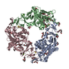
| ||||||||
|---|---|---|---|---|---|---|---|---|---|
| 1 |
| ||||||||
| Unit cell |
|
-Components
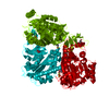
-Protein , 1 types, 3 molecules ABC
| #1: Protein | Mass: 44910.738 Da / Num. of mol.: 3 Source method: isolated from a genetically manipulated source Source: (gene. exp.)  Janthinobacterium sp. (strain J3) (bacteria) Janthinobacterium sp. (strain J3) (bacteria)Strain: J3 / Gene: carAa / Production host: |
|---|
-Non-polymers , 12 types, 896 molecules 


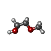


| #2: Chemical |  Mass: 175.820 Da / Num. of mol.: 3 / Source method: obtained synthetically / Formula: Fe2S2 Mass: 175.820 Da / Num. of mol.: 3 / Source method: obtained synthetically / Formula: Fe2S2#3: Chemical |  Mass: 55.845 Da / Num. of mol.: 3 / Source method: obtained synthetically / Formula: Fe / Feature type: SUBJECT OF INVESTIGATION Mass: 55.845 Da / Num. of mol.: 3 / Source method: obtained synthetically / Formula: Fe / Feature type: SUBJECT OF INVESTIGATION#4: Chemical | ChemComp-MG /  Mass: 24.305 Da / Num. of mol.: 5 / Source method: obtained synthetically / Formula: Mg Mass: 24.305 Da / Num. of mol.: 5 / Source method: obtained synthetically / Formula: Mg#5: Chemical | ChemComp-PEG /  Mass: 106.120 Da / Num. of mol.: 8 / Source method: obtained synthetically / Formula: C4H10O3 Mass: 106.120 Da / Num. of mol.: 8 / Source method: obtained synthetically / Formula: C4H10O3#6: Chemical | ChemComp-PGE /  Mass: 150.173 Da / Num. of mol.: 4 / Source method: obtained synthetically / Formula: C6H14O4 Mass: 150.173 Da / Num. of mol.: 4 / Source method: obtained synthetically / Formula: C6H14O4#7: Chemical | ChemComp-PG4 / |  Mass: 194.226 Da / Num. of mol.: 1 / Source method: obtained synthetically / Formula: C8H18O5 / Comment: precipitant*YM Mass: 194.226 Da / Num. of mol.: 1 / Source method: obtained synthetically / Formula: C8H18O5 / Comment: precipitant*YM#8: Chemical | ChemComp-P6G / |  Mass: 282.331 Da / Num. of mol.: 1 / Source method: obtained synthetically / Formula: C12H26O7 / Comment: precipitant*YM Mass: 282.331 Da / Num. of mol.: 1 / Source method: obtained synthetically / Formula: C12H26O7 / Comment: precipitant*YM#9: Chemical |  Mass: 238.278 Da / Num. of mol.: 3 / Source method: obtained synthetically / Formula: C10H22O6 / Comment: precipitant*YM Mass: 238.278 Da / Num. of mol.: 3 / Source method: obtained synthetically / Formula: C10H22O6 / Comment: precipitant*YM#10: Chemical |  Mass: 60.009 Da / Num. of mol.: 2 / Source method: obtained synthetically / Formula: CO3 Mass: 60.009 Da / Num. of mol.: 2 / Source method: obtained synthetically / Formula: CO3#11: Chemical |  Mass: 76.094 Da / Num. of mol.: 2 / Source method: obtained synthetically / Formula: C3H8O2 Mass: 76.094 Da / Num. of mol.: 2 / Source method: obtained synthetically / Formula: C3H8O2#12: Chemical | ChemComp-PG0 /  Mass: 120.147 Da / Num. of mol.: 4 / Source method: obtained synthetically / Formula: C5H12O3 / Comment: precipitant*YM Mass: 120.147 Da / Num. of mol.: 4 / Source method: obtained synthetically / Formula: C5H12O3 / Comment: precipitant*YM#13: Water | ChemComp-HOH / | Mass: 18.015 Da / Num. of mol.: 860 / Source method: isolated from a natural source / Formula: H2O |
|---|
-Details
| Has ligand of interest | Y |
|---|
-Experimental details
-Experiment
| Experiment | Method: X-RAY DIFFRACTION / Number of used crystals: 1 |
|---|
- Sample preparation
| Crystal | Density Matthews: 2.2 Å3/Da / Density % sol: 44.21 % |
|---|---|
| Crystal grow | Temperature: 293 K / Method: vapor diffusion, hanging drop / pH: 7.5 Details: 0.05 M MgCl2, 0.1 M HEPES pH 7.5, 30% (v/v) PEG MME 550 |
-Data collection
| Diffraction | Mean temperature: 100 K / Serial crystal experiment: N |
|---|---|
| Diffraction source | Source: SYNCHROTRON / Site: Photon Factory / Beamline: BL-5A / Wavelength: 1 Å |
| Detector | Type: ADSC QUANTUM 315 / Detector: CCD / Date: Nov 22, 2012 |
| Radiation | Protocol: SINGLE WAVELENGTH / Monochromatic (M) / Laue (L): M / Scattering type: x-ray |
| Radiation wavelength | Wavelength: 1 Å / Relative weight: 1 |
| Reflection | Resolution: 1.6→100 Å / Num. obs: 152740 / % possible obs: 100 % / Redundancy: 21.7 % / Rmerge(I) obs: 0.122 / Net I/σ(I): 18.7 |
| Reflection shell | Resolution: 1.6→1.63 Å / Rmerge(I) obs: 1.576 / Num. unique obs: 7547 |
- Processing
| Software |
| ||||||||||||||||||||||||||||||||||||||||||||||||||||||||||||
|---|---|---|---|---|---|---|---|---|---|---|---|---|---|---|---|---|---|---|---|---|---|---|---|---|---|---|---|---|---|---|---|---|---|---|---|---|---|---|---|---|---|---|---|---|---|---|---|---|---|---|---|---|---|---|---|---|---|---|---|---|---|
| Refinement | Method to determine structure: MOLECULAR REPLACEMENT Starting model: 1WW9 Resolution: 1.6→46.01 Å / Cor.coef. Fo:Fc: 0.972 / Cor.coef. Fo:Fc free: 0.959 / SU B: 1.709 / SU ML: 0.058 / Cross valid method: THROUGHOUT / σ(F): 0 / ESU R: 0.08 / ESU R Free: 0.082 Details: HYDROGENS HAVE BEEN ADDED IN THE RIDING POSITIONS U VALUES : REFINED INDIVIDUALLY
| ||||||||||||||||||||||||||||||||||||||||||||||||||||||||||||
| Solvent computation | Ion probe radii: 0.8 Å / Shrinkage radii: 0.8 Å / VDW probe radii: 1.2 Å | ||||||||||||||||||||||||||||||||||||||||||||||||||||||||||||
| Displacement parameters | Biso max: 77.56 Å2 / Biso mean: 21.3 Å2 / Biso min: 8.23 Å2
| ||||||||||||||||||||||||||||||||||||||||||||||||||||||||||||
| Refinement step | Cycle: final / Resolution: 1.6→46.01 Å
| ||||||||||||||||||||||||||||||||||||||||||||||||||||||||||||
| Refine LS restraints |
| ||||||||||||||||||||||||||||||||||||||||||||||||||||||||||||
| LS refinement shell | Resolution: 1.6→1.642 Å / Rfactor Rfree error: 0 / Total num. of bins used: 20
|
