 Movie
Movie Controller

[English] 日本語
 Yorodumi
- PDB-7boo: Crystal Structure of Core-mannan synthase A (CmsA/Ktr4) from Aspe... -
+ Open data

- Basic information
| Entry | Database: PDB / ID: 7boo | ||||||
|---|---|---|---|---|---|---|---|
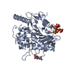
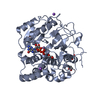
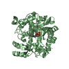
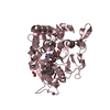
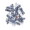
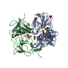
| Title | Crystal Structure of Core-mannan synthase A (CmsA/Ktr4) from Aspergillus fumigatus, apo form | ||||||
 Components Components | Alpha-1,2-mannosyltransferase (Ktr4), putative | ||||||
 Keywords Keywords | TRANSFERASE / Mannosyltransferase | ||||||
| Function / homology |  Function and homology information Function and homology informationalpha-1,2-mannosyltransferase activity / cell wall mannoprotein biosynthetic process / protein O-linked glycosylation / protein N-linked glycosylation / Golgi apparatus / membrane / metal ion binding Similarity search - Function | ||||||
| Biological species |  | ||||||
| Method |  X-RAY DIFFRACTION / SYNCHROTRON / MOLECULAR REPLACEMENT / Resolution: 1.95 Å X-RAY DIFFRACTION / SYNCHROTRON / MOLECULAR REPLACEMENT / Resolution: 1.95 Å | ||||||
 Authors Authors | Hira, D. / Onoue, T. / Oka, T. | ||||||
 Citation Citation | Journal: J.Biol.Chem. / Year: 2020 Title: Structural basis for the core-mannan biosynthesis of cell wall fungal-type galactomannan in Aspergillus fumigatus . Authors: Hira, D. / Onoue, T. / Oka, T. | ||||||
| History |
|
- Structure visualization
| Structure viewer | Molecule: MolmilJmol/JSmol |
|---|
- Downloads & links
-Download
| PDBx/mmCIF format | 7boo.cif.gz | 504.1 KB | Display | PDBx/mmCIF format |
|---|---|---|---|---|
| PDB format | pdb7boo.ent.gz | 412.8 KB | Display | PDB format |
| PDBx/mmJSON format | 7boo.json.gz | Tree view | PDBx/mmJSON format | |
| Others |  Other downloads Other downloads |
-Validation report
| Arichive directory | https://data.pdbj.org/pub/pdb/validation_reports/bo/7booftp://data.pdbj.org/pub/pdb/validation_reports/bo/7boo | HTTPS FTP |
|---|
-Related structure data
| Related structure data |  7bopC 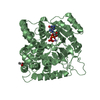 5a07S S: Starting model for refinement C: citing same article ( |
|---|---|
| Similar structure data |


-Links
 PDBj
PDBj

- Assembly
| Deposited unit | 
| ||||||||
|---|---|---|---|---|---|---|---|---|---|
| 1 | 
| ||||||||
| 2 | 
| ||||||||
| 3 | 
| ||||||||
| 4 | 
| ||||||||
| 5 | 
| ||||||||
| 6 | 
| ||||||||
| Unit cell |
| ||||||||
| Components on special symmetry positions |
|
-Components
| #1: Protein | Mass: 45301.371 Da / Num. of mol.: 6 Source method: isolated from a genetically manipulated source Source: (gene. exp.) Strain: CEA10 / CBS 144.89 / FGSC A1163 / Gene: AFUB_051270 / Production host:  #2: Chemical | ChemComp-NA /   Mass: 22.990 Da / Num. of mol.: 7 / Source method: isolated from a natural source / Formula: Na Mass: 22.990 Da / Num. of mol.: 7 / Source method: isolated from a natural source / Formula: Na#3: Chemical | ChemComp-GOL /   Mass: 92.094 Da / Num. of mol.: 21 / Source method: obtained synthetically / Formula: C3H8O3 Mass: 92.094 Da / Num. of mol.: 21 / Source method: obtained synthetically / Formula: C3H8O3#4: Water | ChemComp-HOH / |  Mass: 18.015 Da / Num. of mol.: 1738 / Source method: isolated from a natural source / Formula: H2O Mass: 18.015 Da / Num. of mol.: 1738 / Source method: isolated from a natural source / Formula: H2OHas ligand of interest | N | Has protein modification | Y | |
|---|
-Experimental details
-Experiment
| Experiment | Method: X-RAY DIFFRACTION / Number of used crystals: 1 |
|---|
- Sample preparation
| Crystal | Density Matthews: 3.78 Å3/Da / Density % sol: 67.5 % |
|---|---|
| Crystal grow | Temperature: 277 K / Method: vapor diffusion, hanging drop / Details: sodium citrate |
-Data collection
| Diffraction | Mean temperature: 100 K / Serial crystal experiment: N |
|---|---|
| Diffraction source | Source: SYNCHROTRON / Site: SPring-8  / Beamline: BL44XU / Wavelength: 0.9 Å / Beamline: BL44XU / Wavelength: 0.9 Å |
| Detector | Type: DECTRIS EIGER X 16M / Detector: PIXEL / Date: Oct 20, 2018 |
| Radiation | Protocol: SINGLE WAVELENGTH / Monochromatic (M) / Laue (L): M / Scattering type: x-ray |
| Radiation wavelength | Wavelength: 0.9 Å / Relative weight: 1 |
| Reflection | Resolution: 1.95→45.99 Å / Num. obs: 284394 / % possible obs: 99.79 % / Redundancy: 7 % / CC1/2: 0.999 / Rmerge(I) obs: 0.062 / Rrim(I) all: 0.067 / Net I/σ(I): 19.22 |
| Reflection shell | Resolution: 1.95→2.02 Å / Rmerge(I) obs: 0.555 / Mean I/σ(I) obs: 3.38 / Num. unique obs: 27979 / CC1/2: 0.897 / Rrim(I) all: 0.6 |
- Processing
| Software |
| ||||||||||||||||||||||||||||||||||||||||||||||||||||||||||||||||||||||||||||||||||||||||||||||||||||||||||||||||||||||||||||||||||||||||||||||||||||||||||||||||||||||||||||||||||||||
|---|---|---|---|---|---|---|---|---|---|---|---|---|---|---|---|---|---|---|---|---|---|---|---|---|---|---|---|---|---|---|---|---|---|---|---|---|---|---|---|---|---|---|---|---|---|---|---|---|---|---|---|---|---|---|---|---|---|---|---|---|---|---|---|---|---|---|---|---|---|---|---|---|---|---|---|---|---|---|---|---|---|---|---|---|---|---|---|---|---|---|---|---|---|---|---|---|---|---|---|---|---|---|---|---|---|---|---|---|---|---|---|---|---|---|---|---|---|---|---|---|---|---|---|---|---|---|---|---|---|---|---|---|---|---|---|---|---|---|---|---|---|---|---|---|---|---|---|---|---|---|---|---|---|---|---|---|---|---|---|---|---|---|---|---|---|---|---|---|---|---|---|---|---|---|---|---|---|---|---|---|---|---|---|
| Refinement | Method to determine structure: MOLECULAR REPLACEMENT Starting model: 5a07 Resolution: 1.95→45.99 Å / Cor.coef. Fo:Fc: 0.97 / Cor.coef. Fo:Fc free: 0.962 / SU B: 2.578 / SU ML: 0.071 / Cross valid method: THROUGHOUT / ESU R: 0.107 / ESU R Free: 0.102 / Details: HYDROGENS HAVE BEEN ADDED IN THE RIDING POSITIONS
| ||||||||||||||||||||||||||||||||||||||||||||||||||||||||||||||||||||||||||||||||||||||||||||||||||||||||||||||||||||||||||||||||||||||||||||||||||||||||||||||||||||||||||||||||||||||
| Solvent computation | Ion probe radii: 0.8 Å / Shrinkage radii: 0.8 Å / VDW probe radii: 1.2 Å | ||||||||||||||||||||||||||||||||||||||||||||||||||||||||||||||||||||||||||||||||||||||||||||||||||||||||||||||||||||||||||||||||||||||||||||||||||||||||||||||||||||||||||||||||||||||
| Displacement parameters | Biso mean: 35.973 Å2
| ||||||||||||||||||||||||||||||||||||||||||||||||||||||||||||||||||||||||||||||||||||||||||||||||||||||||||||||||||||||||||||||||||||||||||||||||||||||||||||||||||||||||||||||||||||||
| Refinement step | Cycle: 1 / Resolution: 1.95→45.99 Å
| ||||||||||||||||||||||||||||||||||||||||||||||||||||||||||||||||||||||||||||||||||||||||||||||||||||||||||||||||||||||||||||||||||||||||||||||||||||||||||||||||||||||||||||||||||||||
| Refine LS restraints |
|