 Movie
Movie Controller

[English] 日本語
 Yorodumi
- PDB-7bkr: Endothiapepsin structure obtained at 298K and 40 mM DMSO from a d... -
+ Open data

- Basic information
| Entry | Database: PDB / ID: 7bkr | ||||||
|---|---|---|---|---|---|---|---|
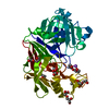
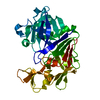
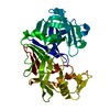
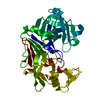
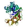
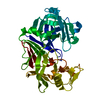
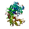
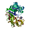
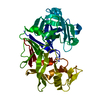
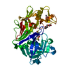
| Title | Endothiapepsin structure obtained at 298K and 40 mM DMSO from a dataset collected with JUNGFRAU detector | ||||||
 Components Components | Endothiapepsin | ||||||
 Keywords Keywords | HYDROLASE / FBDD / DMSO | ||||||
| Function / homology |  Function and homology information Function and homology information | ||||||
| Biological species |  Cryphonectria parasitica (chestnut blight fungus) Cryphonectria parasitica (chestnut blight fungus) | ||||||
| Method |  X-RAY DIFFRACTION / SYNCHROTRON / MOLECULAR REPLACEMENT / molecular replacement / Resolution: 2.1 Å X-RAY DIFFRACTION / SYNCHROTRON / MOLECULAR REPLACEMENT / molecular replacement / Resolution: 2.1 Å | ||||||
 Authors Authors | Engilberge, S. / Huang, C.-Y. / Leonarski, F. / Wojdyla, J.A. / Marsh, M. / Olieric, V. / Wang, M. | ||||||
| Funding support |  Switzerland, 1items Switzerland, 1items
| ||||||
 Citation Citation | Journal: To Be Published Title: Endothiapepsin structure obtained at 298K and 40 mM DMSO from a dataset collected with JUNGFRAU detector Authors: Engilberge, S. / Huang, C.-Y. / Smith, K.M.L. / Eris, D. / Wojdyla, J.A. / Olieric, V. / Leonarski, F. / Sharpe, M. / Wang, M. | ||||||
| History |
|
- Structure visualization
| Structure viewer | Molecule: MolmilJmol/JSmol |
|---|
- Downloads & links
-Download
| PDBx/mmCIF format | 7bkr.cif.gz | 142.7 KB | Display | PDBx/mmCIF format |
|---|---|---|---|---|
| PDB format | pdb7bkr.ent.gz | 110.3 KB | Display | PDB format |
| PDBx/mmJSON format | 7bkr.json.gz | Tree view | PDBx/mmJSON format | |
| Others |  Other downloads Other downloads |
-Validation report
| Arichive directory | https://data.pdbj.org/pub/pdb/validation_reports/bk/7bkrftp://data.pdbj.org/pub/pdb/validation_reports/bk/7bkr | HTTPS FTP |
|---|
-Related structure data
| Related structure data |  6rsvS S: Starting model for refinement |
|---|---|
| Similar structure data |


-Links
 PDBj
PDBj
- Assembly
| Deposited unit | 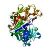
| ||||||||
|---|---|---|---|---|---|---|---|---|---|
| 1 |
| ||||||||
| Unit cell |
|
-Components
| #1: Protein | Mass: 33813.855 Da / Num. of mol.: 1 / Source method: isolated from a natural source Source: (natural) Cryphonectria parasitica (chestnut blight fungus)References: UniProt: P11838, endothiapepsin | ||||||||||
|---|---|---|---|---|---|---|---|---|---|---|---|
| #2: Chemical | ChemComp-DMS /   Mass: 78.133 Da / Num. of mol.: 8 / Source method: obtained synthetically / Formula: C2H6OS / Comment: DMSO, precipitant*YM Mass: 78.133 Da / Num. of mol.: 8 / Source method: obtained synthetically / Formula: C2H6OS / Comment: DMSO, precipitant*YM#3: Chemical | ChemComp-PEG / |   Mass: 106.120 Da / Num. of mol.: 1 / Source method: obtained synthetically / Formula: C4H10O3 Mass: 106.120 Da / Num. of mol.: 1 / Source method: obtained synthetically / Formula: C4H10O3#4: Chemical | ChemComp-1PE / |   Mass: 238.278 Da / Num. of mol.: 1 / Source method: obtained synthetically / Formula: C10H22O6 / Comment: precipitant*YM Mass: 238.278 Da / Num. of mol.: 1 / Source method: obtained synthetically / Formula: C10H22O6 / Comment: precipitant*YM#5: Water | ChemComp-HOH / |  Mass: 18.015 Da / Num. of mol.: 152 / Source method: isolated from a natural source / Formula: H2O Mass: 18.015 Da / Num. of mol.: 152 / Source method: isolated from a natural source / Formula: H2OHas ligand of interest | N | Has protein modification | Y | |
-Experimental details
-Experiment
| Experiment | Method: X-RAY DIFFRACTION / Number of used crystals: 1 |
|---|
- Sample preparation
| Crystal | Density Matthews: 2.51 Å3/Da / Density % sol: 50.95 % |
|---|---|
| Crystal grow | Temperature: 293 K / Method: vapor diffusion Details: 100 mM ammonium acetate, 100 mM sodium acetate pH 4.6 and 26 to 30% PEG 4000 |
-Data collection
| Diffraction | Mean temperature: 298 K / Serial crystal experiment: N |
|---|---|
| Diffraction source | Source: SYNCHROTRON / Site: SLS / Beamline: X06DA / Wavelength: 1 Å |
| Detector | Type: PSI JUNGFRAU 4M / Detector: PIXEL / Date: Oct 24, 2020 |
| Radiation | Protocol: SINGLE WAVELENGTH / Monochromatic (M) / Laue (L): M / Scattering type: x-ray |
| Radiation wavelength | Wavelength: 1 Å / Relative weight: 1 |
| Reflection | Resolution: 2.1→43.33 Å / Num. obs: 19537 / % possible obs: 99.6 % / Redundancy: 5.6 % / CC1/2: 0.97 / Rpim(I) all: 0.2427 / Net I/σ(I): 6.65 |
| Reflection shell | Resolution: 2.1→2.17 Å / Mean I/σ(I) obs: 1.56 / Num. unique obs: 1969 / CC1/2: 0.63 / Rpim(I) all: 0.8456 |
-Phasing
| Phasing | Method: molecular replacement |
|---|
- Processing
| Software |
| ||||||||||||||||||||||||||||||||||||||||||||||||||||||||
|---|---|---|---|---|---|---|---|---|---|---|---|---|---|---|---|---|---|---|---|---|---|---|---|---|---|---|---|---|---|---|---|---|---|---|---|---|---|---|---|---|---|---|---|---|---|---|---|---|---|---|---|---|---|---|---|---|---|
| Refinement | Method to determine structure: MOLECULAR REPLACEMENT Starting model: 6RSV Resolution: 2.1→43.33 Å / SU ML: 0.23 / Cross valid method: THROUGHOUT / σ(F): 1.36 / Phase error: 20.99 / Stereochemistry target values: ML
| ||||||||||||||||||||||||||||||||||||||||||||||||||||||||
| Solvent computation | Shrinkage radii: 0.9 Å / VDW probe radii: 1.11 Å / Solvent model: FLAT BULK SOLVENT MODEL | ||||||||||||||||||||||||||||||||||||||||||||||||||||||||
| Displacement parameters | Biso max: 121.65 Å2 / Biso mean: 39.3817 Å2 / Biso min: 18.32 Å2 | ||||||||||||||||||||||||||||||||||||||||||||||||||||||||
| Refinement step | Cycle: final / Resolution: 2.1→43.33 Å
| ||||||||||||||||||||||||||||||||||||||||||||||||||||||||
| LS refinement shell | Refine-ID: X-RAY DIFFRACTION / Rfactor Rfree error: 0 / Total num. of bins used: 7
| ||||||||||||||||||||||||||||||||||||||||||||||||||||||||
| Refinement TLS params. | Method: refined / Origin x: 3.7454 Å / Origin y: -0.8974 Å / Origin z: 5.6741 Å
| ||||||||||||||||||||||||||||||||||||||||||||||||||||||||
| Refinement TLS group |
|
