Movie
Movie Controller


+ Open data

- Basic information
| Entry | Database: PDB / ID: 7bcw | |||||||||||||||||||||||||||||||||||||||||||||
|---|---|---|---|---|---|---|---|---|---|---|---|---|---|---|---|---|---|---|---|---|---|---|---|---|---|---|---|---|---|---|---|---|---|---|---|---|---|---|---|---|---|---|---|---|---|---|
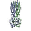
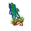
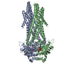
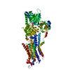
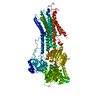
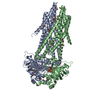
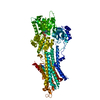
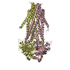
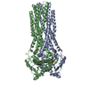
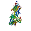
| Title | Structure of MsbA in Salipro with ADP vanadate | |||||||||||||||||||||||||||||||||||||||||||||
 Components Components | ATP-dependent lipid A-core flippase | |||||||||||||||||||||||||||||||||||||||||||||
 Keywords Keywords | MEMBRANE PROTEIN / Lipid export MsbA | |||||||||||||||||||||||||||||||||||||||||||||
| Function / homology |  Function and homology information Function and homology informationMsbA transporter complex / lipopolysaccharide floppase activity / lipid translocation / ABC-type lipid A-core oligosaccharide transporter / lipopolysaccharide transport / ATPase-coupled lipid transmembrane transporter activity / ABC-type xenobiotic transporter activity / lipid transport / ATP-binding cassette (ABC) transporter complex / transmembrane transport ...MsbA transporter complex / lipopolysaccharide floppase activity / lipid translocation / ABC-type lipid A-core oligosaccharide transporter / lipopolysaccharide transport / ATPase-coupled lipid transmembrane transporter activity / ABC-type xenobiotic transporter activity / lipid transport / ATP-binding cassette (ABC) transporter complex / transmembrane transport / lipid binding / ATP hydrolysis activity / ATP binding / membrane / identical protein binding / plasma membrane Similarity search - Function | |||||||||||||||||||||||||||||||||||||||||||||
| Biological species |  | |||||||||||||||||||||||||||||||||||||||||||||
| Method | ELECTRON MICROSCOPY / single particle reconstruction / cryo EM / Resolution: 3.5 Å | |||||||||||||||||||||||||||||||||||||||||||||
 Authors Authors | Traore, D.A.K. / Tidow, H. | |||||||||||||||||||||||||||||||||||||||||||||
 Citation Citation | Journal: FEBS J / Year: 2022 Title: Cryo-EM structure of MsbA in saposin-lipid nanoparticles (Salipro) provides insights into nucleotide coordination. Authors: Dominique-Maurice Kehlenbeck / Daouda A K Traore / Inokentijs Josts / Simon Sander / Martine Moulin / Michael Haertlein / Sylvain Prevost / V Trevor Forsyth / Henning Tidow /    Abstract: The ATP-binding cassette transporter MsbA is a lipid flippase, translocating lipid A, glycolipids, and lipopolysaccharides from the inner to the outer leaflet of the inner membrane of Gram-negative ...The ATP-binding cassette transporter MsbA is a lipid flippase, translocating lipid A, glycolipids, and lipopolysaccharides from the inner to the outer leaflet of the inner membrane of Gram-negative bacteria. It has been used as a model system for time-resolved structural studies as several MsbA structures in different states and reconstitution systems (detergent/nanodiscs/peptidiscs) are available. However, due to the limited resolution of the available structures, detailed structural information on the bound nucleotides has remained elusive. Here, we have reconstituted MsbA in saposin A-lipoprotein nanoparticles (Salipro) and determined the structure of ADP-vanadate-bound MsbA by single-particle cryo-electron microscopy to 3.5 Å resolution. This procedure has resulted in significantly improved resolution and enabled us to model all side chains and visualise detailed ADP-vanadate interactions in the nucleotide-binding domains. The approach may be applicable to other dynamic membrane proteins. | |||||||||||||||||||||||||||||||||||||||||||||
| History |
|
- Structure visualization
| Movie |
Movie viewer |
|---|---|
| Structure viewer | Molecule: MolmilJmol/JSmol |
- Downloads & links
-Download
| PDBx/mmCIF format | 7bcw.cif.gz | 209.2 KB | Display | PDBx/mmCIF format |
|---|---|---|---|---|
| PDB format | pdb7bcw.ent.gz | 164.7 KB | Display | PDB format |
| PDBx/mmJSON format | 7bcw.json.gz | Tree view | PDBx/mmJSON format | |
| Others |  Other downloads Other downloads |
-Validation report
| Arichive directory | https://data.pdbj.org/pub/pdb/validation_reports/bc/7bcwftp://data.pdbj.org/pub/pdb/validation_reports/bc/7bcw | HTTPS FTP |
|---|
-Related structure data
| Related structure data |  12145MC M: map data used to model this data C: citing same article ( |
|---|---|
| Similar structure data |

-Links
 PDBj
PDBj

- Assembly
| Deposited unit | 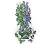
|
|---|---|
| 1 |
|
-Components
| #1: Protein | Mass: 66486.664 Da / Num. of mol.: 2 Source method: isolated from a genetically manipulated source Source: (gene. exp.) Strain: K12 / Gene: msbA, b0914, JW0897 / Production host: References: UniProt: P60752, ABC-type lipid A-core oligosaccharide transporter #2: Chemical |   Mass: 114.939 Da / Num. of mol.: 2 / Source method: obtained synthetically / Formula: VO4 / Feature type: SUBJECT OF INVESTIGATION Mass: 114.939 Da / Num. of mol.: 2 / Source method: obtained synthetically / Formula: VO4 / Feature type: SUBJECT OF INVESTIGATION#3: Chemical |   Mass: 427.201 Da / Num. of mol.: 2 / Source method: obtained synthetically / Formula: C10H15N5O10P2 / Feature type: SUBJECT OF INVESTIGATION / Comment: ADP, energy-carrying molecule*YM Mass: 427.201 Da / Num. of mol.: 2 / Source method: obtained synthetically / Formula: C10H15N5O10P2 / Feature type: SUBJECT OF INVESTIGATION / Comment: ADP, energy-carrying molecule*YM#4: Chemical |   Mass: 24.305 Da / Num. of mol.: 2 / Source method: obtained synthetically / Formula: Mg Mass: 24.305 Da / Num. of mol.: 2 / Source method: obtained synthetically / Formula: Mg#5: Chemical |   Mass: 760.076 Da / Num. of mol.: 2 / Source method: obtained synthetically / Formula: C42H82NO8P / Comment: phospholipid*YM Mass: 760.076 Da / Num. of mol.: 2 / Source method: obtained synthetically / Formula: C42H82NO8P / Comment: phospholipid*YMHas ligand of interest | Y | Has protein modification | N | |
|---|
-Experimental details
-Experiment
| Experiment | Method: ELECTRON MICROSCOPY |
|---|---|
| EM experiment | Aggregation state: PARTICLE / 3D reconstruction method: single particle reconstruction |
- Sample preparation
| Component | Name: MsbA in Salipro with ADP vanadate / Type: COMPLEX / Entity ID: #1 / Source: RECOMBINANT |
|---|---|
| Source (natural) | Organism: |
| Source (recombinant) | Organism: |
| Buffer solution | pH: 8 |
| Specimen | Conc.: 0.6 mg/ml / Embedding applied: NO / Shadowing applied: NO / Staining applied: NO / Vitrification applied: YES |
| Specimen support | Grid material: COPPER / Grid mesh size: 200 divisions/in. / Grid type: Quantifoil R2/1 |
| Vitrification | Cryogen name: ETHANE |
- Electron microscopy imaging
| Experimental equipment |  Model: Titan Krios / Image courtesy: FEI Company |
|---|---|
| Microscopy | Model: FEI TITAN KRIOS |
| Electron gun | Electron source:  FIELD EMISSION GUN / Accelerating voltage: 300 kV / Illumination mode: FLOOD BEAM FIELD EMISSION GUN / Accelerating voltage: 300 kV / Illumination mode: FLOOD BEAM |
| Electron lens | Mode: BRIGHT FIELD |
| Image recording | Electron dose: 40 e/Å2 / Detector mode: COUNTING / Film or detector model: GATAN K2 SUMMIT (4k x 4k) |
| EM imaging optics | Energyfilter name: GIF Quantum LS / Energyfilter slit width: 20 eV |
- Processing
| Software | Name: PHENIX / Version: 1.18.2_3874: / Classification: refinement | ||||||||||||||||||||||||
|---|---|---|---|---|---|---|---|---|---|---|---|---|---|---|---|---|---|---|---|---|---|---|---|---|---|
| EM software |
| ||||||||||||||||||||||||
| CTF correction | Type: PHASE FLIPPING ONLY | ||||||||||||||||||||||||
| Symmetry | Point symmetry: C2 (2 fold cyclic) | ||||||||||||||||||||||||
| 3D reconstruction | Resolution: 3.5 Å / Resolution method: FSC 0.143 CUT-OFF / Num. of particles: 83278 / Symmetry type: POINT | ||||||||||||||||||||||||
| Atomic model building | Protocol: RIGID BODY FIT / Space: REAL | ||||||||||||||||||||||||
| Atomic model building | PDB-ID: 5TTP Pdb chain-ID: A / Accession code: 5TTP / Source name: PDB / Type: experimental model | ||||||||||||||||||||||||
| Refinement | Cross valid method: NONE Stereochemistry target values: GeoStd + Monomer Library + CDL v1.2 | ||||||||||||||||||||||||
| Displacement parameters | Biso mean: 71.61 Å2 | ||||||||||||||||||||||||
| Refine LS restraints |
|