 Movie
Movie Controller


+ Open data

- Basic information
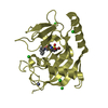
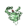
| Entry | Database: PDB / ID: 7bax | ||||||
|---|---|---|---|---|---|---|---|
| Title | Crystal structure of LYS11 ectodomain | ||||||
 Components Components | LysM type receptor kinase | ||||||
 Keywords Keywords | PLANT PROTEIN / LysM-RLK ectodomain | ||||||
| Function / homology |  Function and homology information Function and homology information | ||||||
| Biological species |  | ||||||
| Method |  X-RAY DIFFRACTION / SYNCHROTRON / MOLECULAR REPLACEMENT / Resolution: 2.9 Å X-RAY DIFFRACTION / SYNCHROTRON / MOLECULAR REPLACEMENT / Resolution: 2.9 Å | ||||||
 Authors Authors | Laursen, M. / Cheng, J. / Gysel, K. / Blaise, M. / Andersen, K.R. | ||||||
| Funding support | 1items
| ||||||
 Citation Citation | Journal: Proc.Natl.Acad.Sci.USA / Year: 2021 Title: Kinetic proofreading of lipochitooligosaccharides determines signal activation of symbiotic plant receptors. Authors: Gysel, K. / Laursen, M. / Thygesen, M.B. / Lironi, D. / Bozsoki, Z. / Hjuler, C.T. / Maolanon, N.N. / Cheng, J. / Bjork, P.K. / Vinther, M. / Madsen, L.H. / Rubsam, H. / Muszynski, A. / ...Authors: Gysel, K. / Laursen, M. / Thygesen, M.B. / Lironi, D. / Bozsoki, Z. / Hjuler, C.T. / Maolanon, N.N. / Cheng, J. / Bjork, P.K. / Vinther, M. / Madsen, L.H. / Rubsam, H. / Muszynski, A. / Ghodrati, A. / Azadi, P. / Sullivan, J.T. / Ronson, C.W. / Jensen, K.J. / Blaise, M. / Radutoiu, S. / Stougaard, J. / Andersen, K.R. | ||||||
| History |
|
- Structure visualization
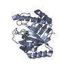
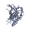
| Structure viewer | Molecule: MolmilJmol/JSmol |
|---|
- Downloads & links
-Download
| PDBx/mmCIF format | 7bax.cif.gz | 109.8 KB | Display | PDBx/mmCIF format |
|---|---|---|---|---|
| PDB format | pdb7bax.ent.gz | 74.2 KB | Display | PDB format |
| PDBx/mmJSON format | 7bax.json.gz | Tree view | PDBx/mmJSON format | |
| Others |  Other downloads Other downloads |
-Validation report
| Arichive directory | https://data.pdbj.org/pub/pdb/validation_reports/ba/7baxftp://data.pdbj.org/pub/pdb/validation_reports/ba/7bax | HTTPS FTP |
|---|
-Related structure data
| Related structure data | 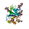 7au7SC S: Starting model for refinement C: citing same article ( |
|---|---|
| Similar structure data |


-Links
 PDBj
PDBj
- Assembly
| Deposited unit | 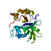
| ||||||||||||
|---|---|---|---|---|---|---|---|---|---|---|---|---|---|
| 1 |
| ||||||||||||
| Unit cell |
|
-Components
| #1: Protein | Mass: 29853.328 Da / Num. of mol.: 1 Source method: isolated from a genetically manipulated source Source: (gene. exp.)   Spodoptera frugiperda (fall armyworm) / References: UniProt: D3KTZ8 Spodoptera frugiperda (fall armyworm) / References: UniProt: D3KTZ8 |
|---|---|
| #2: Sugar | ChemComp-NAG /   Type: D-saccharide, beta linking / Mass: 221.208 Da / Num. of mol.: 1 / Source method: obtained synthetically / Formula: C8H15NO6 Type: D-saccharide, beta linking / Mass: 221.208 Da / Num. of mol.: 1 / Source method: obtained synthetically / Formula: C8H15NO6 |
| Has ligand of interest | N |
| Has protein modification | Y |
-Experimental details
-Experiment
| Experiment | Method: X-RAY DIFFRACTION / Number of used crystals: 1 |
|---|
- Sample preparation
| Crystal | Density Matthews: 4.41 Å3/Da / Density % sol: 72.13 % |
|---|---|
| Crystal grow | Temperature: 277 K / Method: vapor diffusion, sitting drop / pH: 6 / Details: 0.1 M Sodium malonate pH 6.0, 12% PEG3350 |
-Data collection
| Diffraction | Mean temperature: 100 K / Serial crystal experiment: N |
|---|---|
| Diffraction source | Source: SYNCHROTRON / Site: MAX IV  / Beamline: BioMAX / Wavelength: 0.954 Å / Beamline: BioMAX / Wavelength: 0.954 Å |
| Detector | Type: DECTRIS EIGER X 16M / Detector: PIXEL / Date: Feb 22, 2019 |
| Radiation | Monochromator: M / Protocol: SINGLE WAVELENGTH / Monochromatic (M) / Laue (L): M / Scattering type: x-ray |
| Radiation wavelength | Wavelength: 0.954 Å / Relative weight: 1 |
| Reflection | Resolution: 2.88→40 Å / Num. obs: 8086 / % possible obs: 77.1 % / Redundancy: 9.7 % / Biso Wilson estimate: 87.14 Å2 / CC1/2: 0.999 / Rrim(I) all: 0.088 / Net I/σ(I): 15.97 |
| Reflection shell | Resolution: 2.88→2.95 Å / Redundancy: 7.8 % / Mean I/σ(I) obs: 4.93 / Num. unique obs: 60 / CC1/2: 0.897 / Rrim(I) all: 0.472 / % possible all: 7.7 |
- Processing
| Software |
| |||||||||||||||||||||||||||||||||||||||||||||||||
|---|---|---|---|---|---|---|---|---|---|---|---|---|---|---|---|---|---|---|---|---|---|---|---|---|---|---|---|---|---|---|---|---|---|---|---|---|---|---|---|---|---|---|---|---|---|---|---|---|---|---|
| Refinement | Method to determine structure: MOLECULAR REPLACEMENT Starting model: 7AU7 Resolution: 2.9→38.08 Å / SU ML: 0.2735 / Cross valid method: FREE R-VALUE / σ(F): 1.38 / Phase error: 27.0944 Stereochemistry target values: GeoStd + Monomer Library + CDL v1.2
| |||||||||||||||||||||||||||||||||||||||||||||||||
| Solvent computation | Shrinkage radii: 0.9 Å / VDW probe radii: 1.11 Å / Solvent model: FLAT BULK SOLVENT MODEL | |||||||||||||||||||||||||||||||||||||||||||||||||
| Displacement parameters | Biso mean: 77.1 Å2 | |||||||||||||||||||||||||||||||||||||||||||||||||
| Refinement step | Cycle: LAST / Resolution: 2.9→38.08 Å
| |||||||||||||||||||||||||||||||||||||||||||||||||
| Refine LS restraints |
| |||||||||||||||||||||||||||||||||||||||||||||||||
| LS refinement shell |
| |||||||||||||||||||||||||||||||||||||||||||||||||
| Refinement TLS params. | Method: refined / Origin x: 14.4765223404 Å / Origin y: -48.8526891223 Å / Origin z: -8.57149754329 Å
| |||||||||||||||||||||||||||||||||||||||||||||||||
| Refinement TLS group | Selection details: all |
