 Movie
Movie Controller

[English] 日本語
 Yorodumi
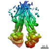
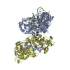
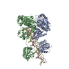
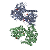
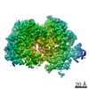
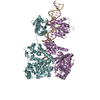
- PDB-7b9q: The SERp optimized structure of Ribonucleotide reductase from Rho... -
+ Open data

- Basic information
| Entry | Database: PDB / ID: 7b9q | ||||||
|---|---|---|---|---|---|---|---|
| Title | The SERp optimized structure of Ribonucleotide reductase from Rhodobacter sphaeroides | ||||||
 Components Components | Vitamin B12-dependent ribonucleotide reductase | ||||||
 Keywords Keywords | OXIDOREDUCTASE / Ribonucleotide Reductase / Thiyl Radical Enzyme / Allosteric Effector | ||||||
| Function / homology | 2'-DEOXYADENOSINE 5'-TRIPHOSPHATE / :  Function and homology information Function and homology information | ||||||
| Biological species |  Rhodobacter sphaeroides (bacteria) Rhodobacter sphaeroides (bacteria) | ||||||
| Method |  X-RAY DIFFRACTION / SYNCHROTRON / MOLECULAR REPLACEMENT / Resolution: 2.78 Å X-RAY DIFFRACTION / SYNCHROTRON / MOLECULAR REPLACEMENT / Resolution: 2.78 Å | ||||||
 Authors Authors | Loderer, C. / Feiler, C. / Wilk, P. / Kabinger, F. | ||||||
 Citation Citation | Journal: Biochemistry / Year: 2022 Title: HUG Domain Is Responsible for Active Dimer Stabilization in an NrdJd Ribonucleotide Reductase. Authors: Fietze, T. / Wilk, P. / Kabinger, F. / Anoosheh, S. / Hofer, A. / Lundin, D. / Feiler, C.G. / Weiss, M.S. / Loderer, C. | ||||||
| History |
|
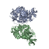
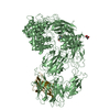
- Structure visualization
| Structure viewer | Molecule: MolmilJmol/JSmol |
|---|
- Downloads & links
-Download
| PDBx/mmCIF format | 7b9q.cif.gz | 683.8 KB | Display | PDBx/mmCIF format |
|---|---|---|---|---|
| PDB format | pdb7b9q.ent.gz | 565.6 KB | Display | PDB format |
| PDBx/mmJSON format | 7b9q.json.gz | Tree view | PDBx/mmJSON format | |
| Others |  Other downloads Other downloads |
-Validation report
| Arichive directory | https://data.pdbj.org/pub/pdb/validation_reports/b9/7b9qftp://data.pdbj.org/pub/pdb/validation_reports/b9/7b9q | HTTPS FTP |
|---|
-Related structure data
| Related structure data |  7b9pSC S: Starting model for refinement C: citing same article ( |
|---|---|
| Similar structure data |
-Links
 PDBj
PDBj


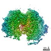
- Assembly
| Deposited unit | 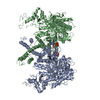
| ||||||||
|---|---|---|---|---|---|---|---|---|---|
| 1 |
| ||||||||
| Unit cell |
|
-Components
| #1: Protein | Mass: 100348.188 Da / Num. of mol.: 2 / Mutation: V926Stop, E96A, E97A, E98A Source method: isolated from a genetically manipulated source Details: From the full length protein with 1218 aa, the C-terminal CRD domain was deleted by insertion of a stop codon at postion 926. In order to reduce the surface entropy, the three glutamic acids ...Details: From the full length protein with 1218 aa, the C-terminal CRD domain was deleted by insertion of a stop codon at postion 926. In order to reduce the surface entropy, the three glutamic acids on the positions 96, 97 and 98 were exchanged to alanine. Source: (gene. exp.) Rhodobacter sphaeroides (bacteria) / Gene: HGN32_08220 / Production host: References: UniProt: A0A6H2IRA4, ribonucleoside-diphosphate reductase #2: Chemical |   Mass: 491.182 Da / Num. of mol.: 2 / Source method: isolated from a natural source / Formula: C10H16N5O12P3 Mass: 491.182 Da / Num. of mol.: 2 / Source method: isolated from a natural source / Formula: C10H16N5O12P3#3: Chemical |   Mass: 24.305 Da / Num. of mol.: 2 / Source method: isolated from a natural source / Formula: Mg Mass: 24.305 Da / Num. of mol.: 2 / Source method: isolated from a natural source / Formula: Mg#4: Water | ChemComp-HOH / |  Mass: 18.015 Da / Num. of mol.: 209 / Source method: isolated from a natural source / Formula: H2O Mass: 18.015 Da / Num. of mol.: 209 / Source method: isolated from a natural source / Formula: H2OHas ligand of interest | N | |
|---|
-Experimental details
-Experiment
| Experiment | Method: X-RAY DIFFRACTION / Number of used crystals: 1 |
|---|
- Sample preparation
| Crystal | Density Matthews: 3.16 Å3/Da / Density % sol: 61.07 % |
|---|---|
| Crystal grow | Temperature: 291 K / Method: vapor diffusion, hanging drop / pH: 6.5 Details: Precipitant: 40% MPD, 5% PEG 8000, 100 mM MES-Bufffer Protein sample: 25 mg/mL Enzyme + 100 uM dATP Ratio 1:1 |
-Data collection
| Diffraction | Mean temperature: 100 K / Serial crystal experiment: N | ||||||||||||||||||||||||||||||||||||||||||||||||||||||||||||||||||||||||||||||||||||||||||||||||||||
|---|---|---|---|---|---|---|---|---|---|---|---|---|---|---|---|---|---|---|---|---|---|---|---|---|---|---|---|---|---|---|---|---|---|---|---|---|---|---|---|---|---|---|---|---|---|---|---|---|---|---|---|---|---|---|---|---|---|---|---|---|---|---|---|---|---|---|---|---|---|---|---|---|---|---|---|---|---|---|---|---|---|---|---|---|---|---|---|---|---|---|---|---|---|---|---|---|---|---|---|---|---|
| Diffraction source | Source: SYNCHROTRON / Site: BESSY  / Beamline: 14.1 / Wavelength: 0.9184 Å / Beamline: 14.1 / Wavelength: 0.9184 Å | ||||||||||||||||||||||||||||||||||||||||||||||||||||||||||||||||||||||||||||||||||||||||||||||||||||
| Detector | Type: DECTRIS PILATUS 6M / Detector: PIXEL / Date: Jan 26, 2019 | ||||||||||||||||||||||||||||||||||||||||||||||||||||||||||||||||||||||||||||||||||||||||||||||||||||
| Radiation | Protocol: SINGLE WAVELENGTH / Monochromatic (M) / Laue (L): M / Scattering type: x-ray | ||||||||||||||||||||||||||||||||||||||||||||||||||||||||||||||||||||||||||||||||||||||||||||||||||||
| Radiation wavelength | Wavelength: 0.9184 Å / Relative weight: 1 | ||||||||||||||||||||||||||||||||||||||||||||||||||||||||||||||||||||||||||||||||||||||||||||||||||||
| Reflection | Resolution: 2.776→46.588 Å / Num. obs: 63329 / % possible obs: 99.8 % / Redundancy: 7.641 % / Biso Wilson estimate: 71.558 Å2 / CC1/2: 0.997 / Rmerge(I) obs: 0.197 / Rrim(I) all: 0.212 / Χ2: 1.009 / Net I/σ(I): 9.63 | ||||||||||||||||||||||||||||||||||||||||||||||||||||||||||||||||||||||||||||||||||||||||||||||||||||
| Reflection shell | Diffraction-ID: 1
|
- Processing
| Software |
| ||||||||||||||||||||||||||||||||||||||||||||||||||||||||||||||||||||||||||||||||||||||||||||||||||||||||||||||||||||||||||||||||||||||||||||||||||||||||||||||||||||||||||||||||||||||||||||||||||||||||
|---|---|---|---|---|---|---|---|---|---|---|---|---|---|---|---|---|---|---|---|---|---|---|---|---|---|---|---|---|---|---|---|---|---|---|---|---|---|---|---|---|---|---|---|---|---|---|---|---|---|---|---|---|---|---|---|---|---|---|---|---|---|---|---|---|---|---|---|---|---|---|---|---|---|---|---|---|---|---|---|---|---|---|---|---|---|---|---|---|---|---|---|---|---|---|---|---|---|---|---|---|---|---|---|---|---|---|---|---|---|---|---|---|---|---|---|---|---|---|---|---|---|---|---|---|---|---|---|---|---|---|---|---|---|---|---|---|---|---|---|---|---|---|---|---|---|---|---|---|---|---|---|---|---|---|---|---|---|---|---|---|---|---|---|---|---|---|---|---|---|---|---|---|---|---|---|---|---|---|---|---|---|---|---|---|---|---|---|---|---|---|---|---|---|---|---|---|---|---|---|---|---|
| Refinement | Method to determine structure: MOLECULAR REPLACEMENT Starting model: 7B9P Resolution: 2.78→46.588 Å / SU ML: 0.47 / Cross valid method: THROUGHOUT / σ(F): 1.35 / Phase error: 29.64 / Stereochemistry target values: ML
| ||||||||||||||||||||||||||||||||||||||||||||||||||||||||||||||||||||||||||||||||||||||||||||||||||||||||||||||||||||||||||||||||||||||||||||||||||||||||||||||||||||||||||||||||||||||||||||||||||||||||
| Solvent computation | Shrinkage radii: 0.9 Å / VDW probe radii: 1.11 Å / Solvent model: FLAT BULK SOLVENT MODEL | ||||||||||||||||||||||||||||||||||||||||||||||||||||||||||||||||||||||||||||||||||||||||||||||||||||||||||||||||||||||||||||||||||||||||||||||||||||||||||||||||||||||||||||||||||||||||||||||||||||||||
| Displacement parameters | Biso max: 229.08 Å2 / Biso mean: 94.5947 Å2 / Biso min: 26.74 Å2 | ||||||||||||||||||||||||||||||||||||||||||||||||||||||||||||||||||||||||||||||||||||||||||||||||||||||||||||||||||||||||||||||||||||||||||||||||||||||||||||||||||||||||||||||||||||||||||||||||||||||||
| Refinement step | Cycle: final / Resolution: 2.78→46.588 Å
| ||||||||||||||||||||||||||||||||||||||||||||||||||||||||||||||||||||||||||||||||||||||||||||||||||||||||||||||||||||||||||||||||||||||||||||||||||||||||||||||||||||||||||||||||||||||||||||||||||||||||
| LS refinement shell | Refine-ID: X-RAY DIFFRACTION / Rfactor Rfree error: 0
| ||||||||||||||||||||||||||||||||||||||||||||||||||||||||||||||||||||||||||||||||||||||||||||||||||||||||||||||||||||||||||||||||||||||||||||||||||||||||||||||||||||||||||||||||||||||||||||||||||||||||
| Refinement TLS params. | Method: refined / Refine-ID: X-RAY DIFFRACTION
| ||||||||||||||||||||||||||||||||||||||||||||||||||||||||||||||||||||||||||||||||||||||||||||||||||||||||||||||||||||||||||||||||||||||||||||||||||||||||||||||||||||||||||||||||||||||||||||||||||||||||
| Refinement TLS group |
|
