Entry Database : PDB / ID : 7b57Title Crystal structure of CaMKII-actinin complex bound to ADP Alpha-actinin-2 Calcium/calmodulin-dependent protein kinase type II subunit alpha Keywords / / / Function / homology Function Domain/homology Component
/ / / / / / / / / / / / / / / / / / / / / / / / / / / / / / / / / / / / / / / / / / / / / / / / / / / / / / / / / / / / / / / / / / / / / / / / / / / / / / / / / / / / / / / / / / / / / / / / / / / / / / / / / / / / / / / / / / / / / / / / / / / / / / / / / / / / / / / / / / / / / / / / / / / / Biological species Mus musculus (house mouse)Homo sapiens (human)Method / / / Resolution : 1.95 Å Authors Zhu, J. / Gold, M. Journal : To Be Published Title : Crystal structure of CaMKII-actinin complex bound to ADPAuthors : Zhu, J. / Gold, M. History Deposition Dec 3, 2020 Deposition site / Processing site Revision 1.0 Jun 22, 2022 Provider / Type Revision 1.1 Jan 31, 2024 Group / Refinement descriptionCategory / chem_comp_bond / pdbx_initial_refinement_model
Show all Show less
 Movie
Movie Controller


 Open data
 Basic information
 Components
 Keywords
 Function and homology information
 Homo sapiens (human)
 X-RAY DIFFRACTION /
  Authors
 Citation
 Structure visualization
 Downloads & links
 Other downloads
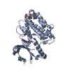
 PDBj
PDBj


 Assembly


 Mass: 24.305 Da / Num. of mol.: 1 / Source method: obtained synthetically / Formula: Mg / Feature type: SUBJECT OF INVESTIGATION
 Mass: 195.237 Da / Num. of mol.: 1 / Source method: obtained synthetically / Formula: C6H13NO4S / Feature type: SUBJECT OF INVESTIGATION / Comment: pH buffer*YM
 Mass: 427.201 Da / Num. of mol.: 1 / Source method: obtained synthetically / Formula: C10H15N5O10P2 / Feature type: SUBJECT OF INVESTIGATION / Comment: ADP, energy-carrying molecule*YM
 Sample preparation
 / Beamline: I04-1 / Wavelength: 0.9119 Å
 Processing