| 登録情報 | データベース: PDB / ID: 7b55
|
|---|
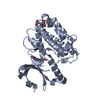
| タイトル | Crystal structure of CaMKII-actinin complex bound to MES |
|---|
 要素 要素 | - Alpha-actinin-2
- Calcium/calmodulin-dependent protein kinase type II subunit alpha
|
|---|
 キーワード キーワード | STRUCTURAL PROTEIN / CaMKII / actinin / dendritic spine |
|---|
| 機能・相同性 |  機能・相同性情報 機能・相同性情報
regulation of synaptic vesicle docking / actin filament uncapping / FATZ binding / titin Z domain binding / HSF1-dependent transactivation / Interferon gamma signaling / positive regulation of endocytic recycling / phospholipase C-activating angiotensin-activated signaling pathway / peptidyl-threonine autophosphorylation / Unblocking of NMDA receptors, glutamate binding and activation ...regulation of synaptic vesicle docking / actin filament uncapping / FATZ binding / titin Z domain binding / HSF1-dependent transactivation / Interferon gamma signaling / positive regulation of endocytic recycling / phospholipase C-activating angiotensin-activated signaling pathway / peptidyl-threonine autophosphorylation / Unblocking of NMDA receptors, glutamate binding and activation / Ion transport by P-type ATPases / RAF activation / calcium- and calmodulin-dependent protein kinase complex / regulation of endocannabinoid signaling pathway / Trafficking of AMPA receptors / Ca2+ pathway / RAF/MAP kinase cascade / positive regulation of cation channel activity / Ca2+/calmodulin-dependent protein kinase / negative regulation of protein localization to cell surface / channel activator activity / LIM domain binding / microspike assembly / dendritic spine development / negative regulation of hydrolase activity / regulation of neurotransmitter secretion / structural constituent of postsynaptic actin cytoskeleton / positive regulation of potassium ion transport / focal adhesion assembly / postsynaptic actin cytoskeleton / muscle cell development / Ion homeostasis / regulation of neuron migration / positive regulation of calcium ion transport / Striated Muscle Contraction / calcium/calmodulin-dependent protein kinase activity / Nephrin family interactions / Assembly and cell surface presentation of NMDA receptors / regulation of mitochondrial membrane permeability involved in apoptotic process / cardiac muscle cell development / structural constituent of muscle / sarcomere organization / cortical actin cytoskeleton / dendrite morphogenesis / pseudopodium / Negative regulation of NMDA receptor-mediated neuronal transmission / Unblocking of NMDA receptors, glutamate binding and activation / negative regulation of potassium ion transport / Long-term potentiation / regulation of neuronal synaptic plasticity / glutamate receptor binding / postsynaptic density, intracellular component / positive regulation of cardiac muscle cell apoptotic process / cellular response to interferon-beta / cytoskeletal protein binding / titin binding / phosphatidylinositol-4,5-bisphosphate binding / Ras activation upon Ca2+ influx through NMDA receptor / platelet alpha granule lumen / response to ischemia / protein localization to plasma membrane / regulation of membrane potential / cell projection / positive regulation of receptor signaling pathway via JAK-STAT / filopodium / actin filament / G1/S transition of mitotic cell cycle / postsynaptic density membrane / Schaffer collateral - CA1 synapse / integrin binding / Z disc / actin filament binding / calcium ion transport / cell junction / Platelet degranulation / RAF/MAP kinase cascade / actin cytoskeleton organization / regulation of apoptotic process / dendritic spine / transmembrane transporter binding / transcription coactivator activity / cytoskeleton / calmodulin binding / cell adhesion / postsynaptic density / protein domain specific binding / protein serine kinase activity / focal adhesion / protein serine/threonine kinase activity / calcium ion binding / synapse / glutamatergic synapse / mitochondrion / extracellular exosome / extracellular region / ATP binding / metal ion binding / identical protein binding / cytosol類似検索 - 分子機能 Calcium/calmodulin-dependent protein kinase II, association-domain / Calcium/calmodulin dependent protein kinase II association domain / EF-hand, Ca insensitive / Ca2+ insensitive EF hand / Ca2+ insensitive EF hand / Spectrin repeat / Spectrin repeat / Actinin-type actin-binding domain signature 1. / Actinin-type actin-binding domain signature 2. / Actinin-type actin-binding domain, conserved site ...Calcium/calmodulin-dependent protein kinase II, association-domain / Calcium/calmodulin dependent protein kinase II association domain / EF-hand, Ca insensitive / Ca2+ insensitive EF hand / Ca2+ insensitive EF hand / Spectrin repeat / Spectrin repeat / Actinin-type actin-binding domain signature 1. / Actinin-type actin-binding domain signature 2. / Actinin-type actin-binding domain, conserved site / Spectrin/alpha-actinin / Spectrin repeats / Calponin homology domain / Calponin homology (CH) domain / Calponin homology domain / CH domain superfamily / Calponin homology (CH) domain profile. / NTF2-like domain superfamily / EF-hand domain pair / EF-hand, calcium binding motif / EF-hand calcium-binding domain profile. / EF-hand domain / EF-hand domain pair / Serine/threonine-protein kinase, active site / Serine/Threonine protein kinases active-site signature. / Protein kinase domain / Serine/Threonine protein kinases, catalytic domain / Protein kinase, ATP binding site / Protein kinases ATP-binding region signature. / Protein kinase domain profile. / Protein kinase domain / Protein kinase-like domain superfamily類似検索 - ドメイン・相同性 Calcium/calmodulin-dependent protein kinase type II subunit alpha / Alpha-actinin-2類似検索 - 構成要素 |
|---|
| 生物種 |   Mus musculus (ハツカネズミ) Mus musculus (ハツカネズミ)
 Homo sapiens (ヒト) Homo sapiens (ヒト) |
|---|
| 手法 |  X線回折 / シンクロトロン / 分子置換 / 解像度: 1.6 Å X線回折 / シンクロトロン / 分子置換 / 解像度: 1.6 Å |
|---|
 データ登録者 データ登録者 | Zhu, J. / Gold, M. |
|---|
 引用 引用 | ジャーナル: To Be Published
タイトル: Crystal structure of CaMKII-actinin complex bound to MES
著者: Zhu, J. / Gold, M. |
|---|
| 履歴 | | 登録 | 2020年12月3日 | 登録サイト: PDBE / 処理サイト: PDBE |
|---|
| 改定 1.0 | 2022年6月22日 | Provider: repository / タイプ: Initial release |
|---|
| 改定 1.1 | 2024年1月31日 | Group: Data collection / Refinement description
カテゴリ: chem_comp_atom / chem_comp_bond / pdbx_initial_refinement_model |
|---|
|
|---|
 ムービー
ムービー コントローラー


 データを開く
 基本情報
基本情報 構造の表示
構造の表示 ダウンロードとリンク
 その他のダウンロード
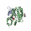
 PDBj
PDBj


 集合体
集合体


 分子量: 195.237 Da / 分子数: 2 / 由来タイプ: 合成 / 式: C6H13NO4S / タイプ: SUBJECT OF INVESTIGATION / コメント: pH緩衝剤*YM
 分子量: 18.015 Da / 分子数: 383 / 由来タイプ: 天然 / 式: H2O
 試料調製
試料調製 / ビームライン: I04-1 / 波長: 0.9119 Å
 解析
解析