+ Open data
- Basic information
| Entry | Database: PDB / ID: 7b55 | ||||||
|---|---|---|---|---|---|---|---|
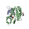
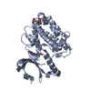
| Title | Crystal structure of CaMKII-actinin complex bound to MES | ||||||
 Components Components |
| ||||||
 Keywords Keywords | STRUCTURAL PROTEIN / CaMKII / actinin / dendritic spine | ||||||
| Function / homology |  Function and homology information Function and homology informationregulation of synaptic vesicle docking / actin filament uncapping / FATZ binding / titin Z domain binding / HSF1-dependent transactivation / Interferon gamma signaling / positive regulation of endocytic recycling / phospholipase C-activating angiotensin-activated signaling pathway / peptidyl-threonine autophosphorylation / Ion transport by P-type ATPases ...regulation of synaptic vesicle docking / actin filament uncapping / FATZ binding / titin Z domain binding / HSF1-dependent transactivation / Interferon gamma signaling / positive regulation of endocytic recycling / phospholipase C-activating angiotensin-activated signaling pathway / peptidyl-threonine autophosphorylation / Ion transport by P-type ATPases / Unblocking of NMDA receptors, glutamate binding and activation / RAF activation / calcium- and calmodulin-dependent protein kinase complex / regulation of endocannabinoid signaling pathway / Trafficking of AMPA receptors / Ca2+ pathway / RAF/MAP kinase cascade / positive regulation of cation channel activity / Ca2+/calmodulin-dependent protein kinase / negative regulation of protein localization to cell surface / channel activator activity / LIM domain binding / microspike assembly / dendritic spine development / negative regulation of hydrolase activity / regulation of neurotransmitter secretion / structural constituent of postsynaptic actin cytoskeleton / positive regulation of potassium ion transport / focal adhesion assembly / muscle cell development / postsynaptic actin cytoskeleton / Ion homeostasis / positive regulation of calcium ion transport / regulation of neuron migration / Striated Muscle Contraction / calcium/calmodulin-dependent protein kinase activity / Nephrin family interactions / Assembly and cell surface presentation of NMDA receptors / regulation of mitochondrial membrane permeability involved in apoptotic process / cardiac muscle cell development / sarcomere organization / structural constituent of muscle / cortical actin cytoskeleton / dendrite morphogenesis / pseudopodium / Negative regulation of NMDA receptor-mediated neuronal transmission / Unblocking of NMDA receptors, glutamate binding and activation / negative regulation of potassium ion transport / Long-term potentiation / regulation of neuronal synaptic plasticity / glutamate receptor binding / postsynaptic density, intracellular component / positive regulation of cardiac muscle cell apoptotic process / cellular response to interferon-beta / cytoskeletal protein binding / titin binding / phosphatidylinositol-4,5-bisphosphate binding / Ras activation upon Ca2+ influx through NMDA receptor / platelet alpha granule lumen / response to ischemia / protein localization to plasma membrane / regulation of membrane potential / cell projection / filopodium / positive regulation of receptor signaling pathway via JAK-STAT / actin filament / G1/S transition of mitotic cell cycle / postsynaptic density membrane / Schaffer collateral - CA1 synapse / integrin binding / Z disc / actin filament binding / cell junction / calcium ion transport / Platelet degranulation / RAF/MAP kinase cascade / actin cytoskeleton organization / regulation of apoptotic process / dendritic spine / transmembrane transporter binding / transcription coactivator activity / cytoskeleton / calmodulin binding / cell adhesion / postsynaptic density / protein domain specific binding / protein serine kinase activity / focal adhesion / protein serine/threonine kinase activity / calcium ion binding / synapse / glutamatergic synapse / mitochondrion / extracellular exosome / extracellular region / ATP binding / metal ion binding / identical protein binding / cytosol Similarity search - Function | ||||||
| Biological species |   Homo sapiens (human) Homo sapiens (human) | ||||||
| Method |  X-RAY DIFFRACTION / X-RAY DIFFRACTION /  SYNCHROTRON / SYNCHROTRON /  MOLECULAR REPLACEMENT / Resolution: 1.6 Å MOLECULAR REPLACEMENT / Resolution: 1.6 Å | ||||||
 Authors Authors | Zhu, J. / Gold, M. | ||||||
 Citation Citation |  Journal: To Be Published Journal: To Be PublishedTitle: Crystal structure of CaMKII-actinin complex bound to MES Authors: Zhu, J. / Gold, M. | ||||||
| History |
|
- Structure visualization
| Structure viewer | Molecule:  Molmil Molmil Jmol/JSmol Jmol/JSmol |
|---|
- Downloads & links
- Download
| PDBx/mmCIF format |  7b55.cif.gz 7b55.cif.gz | 201 KB | Display |  PDBx/mmCIF format PDBx/mmCIF format |
|---|---|---|---|---|
| PDB format |  pdb7b55.ent.gz pdb7b55.ent.gz | 131.2 KB | Display |  PDB format PDB format |
| PDBx/mmJSON format |  7b55.json.gz 7b55.json.gz | Tree view |  PDBx/mmJSON format PDBx/mmJSON format | |
| Others |  Other downloads Other downloads |
-Validation report
| Summary document |  7b55_validation.pdf.gz 7b55_validation.pdf.gz | 1 MB | Display |  wwPDB validaton report wwPDB validaton report |
|---|---|---|---|---|
| Full document |  7b55_full_validation.pdf.gz 7b55_full_validation.pdf.gz | 1 MB | Display | |
| Data in XML |  7b55_validation.xml.gz 7b55_validation.xml.gz | 19.7 KB | Display | |
| Data in CIF |  7b55_validation.cif.gz 7b55_validation.cif.gz | 29.7 KB | Display | |
| Arichive directory |  https://data.pdbj.org/pub/pdb/validation_reports/b5/7b55 https://data.pdbj.org/pub/pdb/validation_reports/b5/7b55 ftp://data.pdbj.org/pub/pdb/validation_reports/b5/7b55 ftp://data.pdbj.org/pub/pdb/validation_reports/b5/7b55 | HTTPS FTP |
-Related structure data
| Related structure data |  7b56C  2vz6S S: Starting model for refinement C: citing same article ( |
|---|---|
| Similar structure data | Similarity search - Function & homology  F&H Search F&H Search |
- Links
Links
- Assembly
| Deposited unit | 
| ||||||||||||
|---|---|---|---|---|---|---|---|---|---|---|---|---|---|
| 1 |
| ||||||||||||
| Unit cell |
|
- Components
| #1: Protein | Mass: 35903.430 Da / Num. of mol.: 1 Source method: isolated from a genetically manipulated source Source: (gene. exp.)   References: UniProt: P11798, Ca2+/calmodulin-dependent protein kinase | ||||
|---|---|---|---|---|---|
| #2: Protein | Mass: 7903.778 Da / Num. of mol.: 1 Source method: isolated from a genetically manipulated source Source: (gene. exp.)  Homo sapiens (human) / Gene: ACTN2 / Production host: Homo sapiens (human) / Gene: ACTN2 / Production host:  | ||||
| #3: Chemical | | #4: Water | ChemComp-HOH / | Has ligand of interest | Y | |
-Experimental details
-Experiment
| Experiment | Method:  X-RAY DIFFRACTION / Number of used crystals: 1 X-RAY DIFFRACTION / Number of used crystals: 1 |
|---|
- Sample preparation
| Crystal | Density Matthews: 2.43 Å3/Da / Density % sol: 49.37 % |
|---|---|
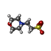
| Crystal grow | Temperature: 277 K / Method: vapor diffusion, sitting drop Details: 0.1 M MES pH 6.0, 20% w/v PEG4000, 0.2 M Lithium Sulfate |
-Data collection
| Diffraction | Mean temperature: 100 K / Serial crystal experiment: N |
|---|---|
| Diffraction source | Source:  SYNCHROTRON / Site: SYNCHROTRON / Site:  Diamond Diamond  / Beamline: I04-1 / Wavelength: 0.9119 Å / Beamline: I04-1 / Wavelength: 0.9119 Å |
| Detector | Type: DECTRIS PILATUS3 S 6M / Detector: PIXEL / Date: Mar 1, 2020 |
| Radiation | Protocol: SINGLE WAVELENGTH / Monochromatic (M) / Laue (L): M / Scattering type: x-ray |
| Radiation wavelength | Wavelength: 0.9119 Å / Relative weight: 1 |
| Reflection | Resolution: 1.6→91.32 Å / Num. obs: 57062 / % possible obs: 100 % / Redundancy: 12.6 % / Biso Wilson estimate: 31.35 Å2 / CC1/2: 1 / Rpim(I) all: 0.018 / Net I/σ(I): 14.6 |
| Reflection shell | Resolution: 1.6→1.63 Å / Mean I/σ(I) obs: 0.8 / Num. unique obs: 2778 / CC1/2: 0.639 / Rpim(I) all: 0.783 |
- Processing
| Software |
| |||||||||||||||||||||||||||||||||||||||||||||||||||||||||||||||||||||||||||||||||||||||||||||||||||||||||
|---|---|---|---|---|---|---|---|---|---|---|---|---|---|---|---|---|---|---|---|---|---|---|---|---|---|---|---|---|---|---|---|---|---|---|---|---|---|---|---|---|---|---|---|---|---|---|---|---|---|---|---|---|---|---|---|---|---|---|---|---|---|---|---|---|---|---|---|---|---|---|---|---|---|---|---|---|---|---|---|---|---|---|---|---|---|---|---|---|---|---|---|---|---|---|---|---|---|---|---|---|---|---|---|---|---|---|
| Refinement | Method to determine structure:  MOLECULAR REPLACEMENT MOLECULAR REPLACEMENTStarting model: 2VZ6 Resolution: 1.6→55.57 Å / SU ML: 0.2062 / Cross valid method: FREE R-VALUE / σ(F): 1.34 / Phase error: 28.8893 Stereochemistry target values: GeoStd + Monomer Library + CDL v1.2
| |||||||||||||||||||||||||||||||||||||||||||||||||||||||||||||||||||||||||||||||||||||||||||||||||||||||||
| Solvent computation | Shrinkage radii: 0.9 Å / VDW probe radii: 1.11 Å / Solvent model: FLAT BULK SOLVENT MODEL | |||||||||||||||||||||||||||||||||||||||||||||||||||||||||||||||||||||||||||||||||||||||||||||||||||||||||
| Displacement parameters | Biso mean: 40.7 Å2 | |||||||||||||||||||||||||||||||||||||||||||||||||||||||||||||||||||||||||||||||||||||||||||||||||||||||||
| Refinement step | Cycle: LAST / Resolution: 1.6→55.57 Å
| |||||||||||||||||||||||||||||||||||||||||||||||||||||||||||||||||||||||||||||||||||||||||||||||||||||||||
| Refine LS restraints |
| |||||||||||||||||||||||||||||||||||||||||||||||||||||||||||||||||||||||||||||||||||||||||||||||||||||||||
| LS refinement shell |
| |||||||||||||||||||||||||||||||||||||||||||||||||||||||||||||||||||||||||||||||||||||||||||||||||||||||||
| Refinement TLS params. | Method: refined / Origin x: 15.9133667869 Å / Origin y: -5.11448531575 Å / Origin z: -2.38755678608 Å
| |||||||||||||||||||||||||||||||||||||||||||||||||||||||||||||||||||||||||||||||||||||||||||||||||||||||||
| Refinement TLS group | Selection details: all |
 Movie
Movie Controller


 PDBj
PDBj


