 Movie
Movie Controller


+ Open data

- Basic information
| Entry | Database: PDB / ID: 7b3n | ||||||
|---|---|---|---|---|---|---|---|
| Title | AmiP amidase-3 from Thermus parvatiensis | ||||||
 Components Components | Cell wall hydrolase | ||||||
 Keywords Keywords | HYDROLASE / amidase-3 thermophilic | ||||||
| Function / homology | : / N-acetylmuramoyl-L-alanine amidase, catalytic domain / N-acetylmuramoyl-L-alanine amidase / N-acetylmuramoyl-L-alanine amidase activity / peptidoglycan catabolic process / outer membrane-bounded periplasmic space / metal ion binding / Cell wall hydrolase Function and homology information Function and homology information | ||||||
| Biological species |   Thermus parvatiensis (bacteria) Thermus parvatiensis (bacteria) | ||||||
| Method |  X-RAY DIFFRACTION / SYNCHROTRON / SAD / Resolution: 1.793 Å X-RAY DIFFRACTION / SYNCHROTRON / SAD / Resolution: 1.793 Å | ||||||
 Authors Authors | Freitag-Pohl, S. / Pohl, E. | ||||||
| Funding support |  United Kingdom, 1items United Kingdom, 1items
| ||||||
 Citation Citation | Journal: Protein Sci. / Year: 2023 Title: AmiP from hyperthermophilic Thermus parvatiensis prophage is a thermoactive and ultrathermostable peptidoglycan lytic amidase. Authors: Jasilionis, A. / Plotka, M. / Wang, L. / Dorawa, S. / Lange, J. / Watzlawick, H. / van den Bergh, T. / Vroling, B. / Altenbuchner, J. / Kaczorowska, A.K. / Pohl, E. / Kaczorowski, T. / ...Authors: Jasilionis, A. / Plotka, M. / Wang, L. / Dorawa, S. / Lange, J. / Watzlawick, H. / van den Bergh, T. / Vroling, B. / Altenbuchner, J. / Kaczorowska, A.K. / Pohl, E. / Kaczorowski, T. / Nordberg Karlsson, E. / Freitag-Pohl, S. | ||||||
| History |
|
- Structure visualization
| Structure viewer | Molecule: MolmilJmol/JSmol |
|---|
- Downloads & links
-Download
| PDBx/mmCIF format | 7b3n.cif.gz | 330.6 KB | Display | PDBx/mmCIF format |
|---|---|---|---|---|
| PDB format | pdb7b3n.ent.gz | 264 KB | Display | PDB format |
| PDBx/mmJSON format | 7b3n.json.gz | Tree view | PDBx/mmJSON format | |
| Others |  Other downloads Other downloads |
-Validation report
| Arichive directory | https://data.pdbj.org/pub/pdb/validation_reports/b3/7b3nftp://data.pdbj.org/pub/pdb/validation_reports/b3/7b3n | HTTPS FTP |
|---|
-Related structure data
| Similar structure data |
|---|
-Links
 PDBj
PDBj- Assembly
| Deposited unit | 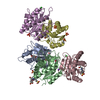
| ||||||||
|---|---|---|---|---|---|---|---|---|---|
| 1 | 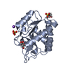
| ||||||||
| 2 | 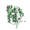
| ||||||||
| 3 | 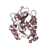
| ||||||||
| 4 | 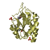
| ||||||||
| 5 | 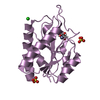
| ||||||||
| Unit cell |
|
-Components
-Protein , 1 types, 5 molecules ABCDE
| #1: Protein | Mass: 19195.770 Da / Num. of mol.: 5 Source method: isolated from a genetically manipulated source Source: (gene. exp.) Thermus parvatiensis (bacteria) / Gene: AV541_09585, RLTM_01600 / Production host: |
|---|
-Non-polymers , 7 types, 274 molecules 


| #2: Chemical | ChemComp-ZN /  Mass: 65.409 Da / Num. of mol.: 5 / Source method: obtained synthetically / Formula: Zn Mass: 65.409 Da / Num. of mol.: 5 / Source method: obtained synthetically / Formula: Zn#3: Chemical | ChemComp-GOL /  Mass: 92.094 Da / Num. of mol.: 5 / Source method: obtained synthetically / Formula: C3H8O3 Mass: 92.094 Da / Num. of mol.: 5 / Source method: obtained synthetically / Formula: C3H8O3#4: Chemical | ChemComp-EPE /  Mass: 238.305 Da / Num. of mol.: 5 / Source method: obtained synthetically / Formula: C8H18N2O4S / Comment: pH buffer*YM Mass: 238.305 Da / Num. of mol.: 5 / Source method: obtained synthetically / Formula: C8H18N2O4S / Comment: pH buffer*YM#5: Chemical | ChemComp-SO4 /  Mass: 96.063 Da / Num. of mol.: 5 / Source method: obtained synthetically / Formula: SO4 Mass: 96.063 Da / Num. of mol.: 5 / Source method: obtained synthetically / Formula: SO4#6: Chemical |  Mass: 22.990 Da / Num. of mol.: 2 / Source method: obtained synthetically / Formula: Na Mass: 22.990 Da / Num. of mol.: 2 / Source method: obtained synthetically / Formula: Na#7: Chemical |  Mass: 35.453 Da / Num. of mol.: 2 / Source method: obtained synthetically / Formula: Cl Mass: 35.453 Da / Num. of mol.: 2 / Source method: obtained synthetically / Formula: Cl#8: Water | ChemComp-HOH / | Mass: 18.015 Da / Num. of mol.: 250 / Source method: isolated from a natural source / Formula: H2O |
|---|
-Details
| Has ligand of interest | N |
|---|
-Experimental details
-Experiment
| Experiment | Method: X-RAY DIFFRACTION / Number of used crystals: 1 |
|---|
- Sample preparation
| Crystal | Density Matthews: 2.45 Å3/Da / Density % sol: 49.79 % |
|---|---|
| Crystal grow | Temperature: 298 K / Method: vapor diffusion, sitting drop / pH: 7.8 / Details: 0.1 M hepes pH 7.8, 65 % MPD |
-Data collection
| Diffraction | Mean temperature: 100 K / Serial crystal experiment: N |
|---|---|
| Diffraction source | Source: SYNCHROTRON / Site: Diamond / Beamline: I03 / Wavelength: 1.2824 Å |
| Detector | Type: DECTRIS EIGER2 XE 16M / Detector: PIXEL / Date: May 3, 2019 |
| Radiation | Protocol: SINGLE WAVELENGTH / Monochromatic (M) / Laue (L): M / Scattering type: x-ray |
| Radiation wavelength | Wavelength: 1.2824 Å / Relative weight: 1 |
| Reflection | Resolution: 1.79→46.53 Å / Num. obs: 86433 / % possible obs: 96.9 % / Redundancy: 1.9 % / CC1/2: 0.999 / Rmerge(I) obs: 0.035 / Rpim(I) all: 0.035 / Rrim(I) all: 0.05 / Χ2: 1.01 / Net I/σ(I): 9.2 |
| Reflection shell | Resolution: 1.79→1.83 Å / Redundancy: 1.9 % / Rmerge(I) obs: 1.132 / Mean I/σ(I) obs: 0.8 / Num. unique obs: 3209 / CC1/2: 0.283 / Rpim(I) all: 1.132 / Rrim(I) all: 1.601 / Χ2: 1.21 / % possible all: 69.3 |
- Processing
| Software |
| ||||||||||||||||||||||||||||||||||||||||||||||||||||||||||||||||||||||||||||||||||||||||||||||||||||||||||||||||||||||||||||||||||||||||||||||||||||||||||||||||
|---|---|---|---|---|---|---|---|---|---|---|---|---|---|---|---|---|---|---|---|---|---|---|---|---|---|---|---|---|---|---|---|---|---|---|---|---|---|---|---|---|---|---|---|---|---|---|---|---|---|---|---|---|---|---|---|---|---|---|---|---|---|---|---|---|---|---|---|---|---|---|---|---|---|---|---|---|---|---|---|---|---|---|---|---|---|---|---|---|---|---|---|---|---|---|---|---|---|---|---|---|---|---|---|---|---|---|---|---|---|---|---|---|---|---|---|---|---|---|---|---|---|---|---|---|---|---|---|---|---|---|---|---|---|---|---|---|---|---|---|---|---|---|---|---|---|---|---|---|---|---|---|---|---|---|---|---|---|---|---|---|---|
| Refinement | Method to determine structure: SAD / Resolution: 1.793→43.679 Å / Cor.coef. Fo:Fc: 0.966 / Cor.coef. Fo:Fc free: 0.951 / SU B: 3.336 / SU ML: 0.096 / Cross valid method: FREE R-VALUE / ESU R: 0.119 / ESU R Free: 0.119 Details: Hydrogens have been added in their riding positions
| ||||||||||||||||||||||||||||||||||||||||||||||||||||||||||||||||||||||||||||||||||||||||||||||||||||||||||||||||||||||||||||||||||||||||||||||||||||||||||||||||
| Solvent computation | Ion probe radii: 0.8 Å / Shrinkage radii: 0.8 Å / VDW probe radii: 1.2 Å / Solvent model: MASK BULK SOLVENT | ||||||||||||||||||||||||||||||||||||||||||||||||||||||||||||||||||||||||||||||||||||||||||||||||||||||||||||||||||||||||||||||||||||||||||||||||||||||||||||||||
| Displacement parameters | Biso mean: 38.893 Å2
| ||||||||||||||||||||||||||||||||||||||||||||||||||||||||||||||||||||||||||||||||||||||||||||||||||||||||||||||||||||||||||||||||||||||||||||||||||||||||||||||||
| Refinement step | Cycle: LAST / Resolution: 1.793→43.679 Å
| ||||||||||||||||||||||||||||||||||||||||||||||||||||||||||||||||||||||||||||||||||||||||||||||||||||||||||||||||||||||||||||||||||||||||||||||||||||||||||||||||
| Refine LS restraints |
| ||||||||||||||||||||||||||||||||||||||||||||||||||||||||||||||||||||||||||||||||||||||||||||||||||||||||||||||||||||||||||||||||||||||||||||||||||||||||||||||||
| LS refinement shell |
|
