 Movie
Movie Controller

[English] 日本語
 Yorodumi
- PDB-7b1x: Crystal structure of cold-active esterase PMGL3 from permafrost m... -
+ Open data

- Basic information
| Entry | Database: PDB / ID: 7b1x | ||||||
|---|---|---|---|---|---|---|---|
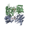
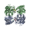
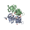
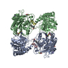
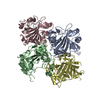
| Title | Crystal structure of cold-active esterase PMGL3 from permafrost metagenomic library | ||||||
 Components Components | esterase PMGL3 | ||||||
 Keywords Keywords | HYDROLASE / esterase / HSL / permafrost / cold-active / psychrophilic / tetramer / dimer / lipase | ||||||
| Function / homology | Alpha/Beta hydrolase fold, catalytic domain / Rossmann fold / 3-Layer(aba) Sandwich / Alpha Beta Function and homology information Function and homology information | ||||||
| Biological species |  uncultured bacterium (environmental samples) uncultured bacterium (environmental samples) | ||||||
| Method |  X-RAY DIFFRACTION / SYNCHROTRON / MOLECULAR REPLACEMENT / molecular replacement / Resolution: 2.3 Å X-RAY DIFFRACTION / SYNCHROTRON / MOLECULAR REPLACEMENT / molecular replacement / Resolution: 2.3 Å | ||||||
 Authors Authors | Boyko, K.M. / Nikolaeva, A.Y. / Petrovskaya, L.E. / Kryukova, M.V. / Kryukova, E.A. / Korzhenevsky, D.A. / Lomakina, G.Y. / Novototskaya-Vlasova, K.A. / Rivkina, E.M. / Dolgikh, D.A. ...Boyko, K.M. / Nikolaeva, A.Y. / Petrovskaya, L.E. / Kryukova, M.V. / Kryukova, E.A. / Korzhenevsky, D.A. / Lomakina, G.Y. / Novototskaya-Vlasova, K.A. / Rivkina, E.M. / Dolgikh, D.A. / Kirpichnikov, M.P. / Popov, V.O. | ||||||
| Funding support |  Russian Federation, 1items Russian Federation, 1items
| ||||||
 Citation Citation | Journal: Biomolecules / Year: 2021 Title: Structural and Biochemical Characterization of a Cold-Active PMGL3 Esterase with Unusual Oligomeric Structure. Authors: Boyko, K.M. / Kryukova, M.V. / Petrovskaya, L.E. / Kryukova, E.A. / Nikolaeva, A.Y. / Korzhenevsky, D.A. / Lomakina, G.Y. / Novototskaya-Vlasova, K.A. / Rivkina, E.M. / Dolgikh, D.A. / ...Authors: Boyko, K.M. / Kryukova, M.V. / Petrovskaya, L.E. / Kryukova, E.A. / Nikolaeva, A.Y. / Korzhenevsky, D.A. / Lomakina, G.Y. / Novototskaya-Vlasova, K.A. / Rivkina, E.M. / Dolgikh, D.A. / Kirpichnikov, M.P. / Popov, V.O. | ||||||
| History |
|
- Structure visualization
| Structure viewer | Molecule: MolmilJmol/JSmol |
|---|
- Downloads & links
-Download
| PDBx/mmCIF format | 7b1x.cif.gz | 237 KB | Display | PDBx/mmCIF format |
|---|---|---|---|---|
| PDB format | pdb7b1x.ent.gz | 190.5 KB | Display | PDB format |
| PDBx/mmJSON format | 7b1x.json.gz | Tree view | PDBx/mmJSON format | |
| Others |  Other downloads Other downloads |
-Validation report
| Arichive directory | https://data.pdbj.org/pub/pdb/validation_reports/b1/7b1xftp://data.pdbj.org/pub/pdb/validation_reports/b1/7b1x | HTTPS FTP |
|---|
-Related structure data
| Related structure data | 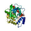 3fakS S: Starting model for refinement |
|---|---|
| Similar structure data |


-Links
 PDBj
PDBj- Assembly
| Deposited unit | 
| ||||||||||||||||||
|---|---|---|---|---|---|---|---|---|---|---|---|---|---|---|---|---|---|---|---|
| 1 | 
| ||||||||||||||||||
| 2 | 
| ||||||||||||||||||
| 3 |
| ||||||||||||||||||
| Unit cell |
| ||||||||||||||||||
| Noncrystallographic symmetry (NCS) | NCS domain:
NCS domain segments: Component-ID: _ / Ens-ID: 1 / Beg auth comp-ID: MET / Beg label comp-ID: MET / End auth comp-ID: GLY / End label comp-ID: GLY / Refine code: _ / Auth seq-ID: 1 - 302 / Label seq-ID: 1 - 302
|
-Components
| #1: Protein | Mass: 33440.672 Da / Num. of mol.: 2 Source method: isolated from a genetically manipulated source Source: (gene. exp.) uncultured bacterium (environmental samples)Production host: #2: Water | ChemComp-HOH / |  Mass: 18.015 Da / Num. of mol.: 197 / Source method: isolated from a natural source / Formula: H2O Mass: 18.015 Da / Num. of mol.: 197 / Source method: isolated from a natural source / Formula: H2O |
|---|
-Experimental details
-Experiment
| Experiment | Method: X-RAY DIFFRACTION / Number of used crystals: 1 |
|---|
- Sample preparation
| Crystal | Density Matthews: 2.26 Å3/Da / Density % sol: 45.56 % |
|---|---|
| Crystal grow | Temperature: 288 K / Method: vapor diffusion Details: 0.1M Sodium citrate tribasic dihydrate pH 5.6; 20% isopropanol; 20% PEG4000 |
-Data collection
| Diffraction | Mean temperature: 100 K / Serial crystal experiment: N | ||||||||||||||||||||||||||||||||||||||||||||||||||||||||
|---|---|---|---|---|---|---|---|---|---|---|---|---|---|---|---|---|---|---|---|---|---|---|---|---|---|---|---|---|---|---|---|---|---|---|---|---|---|---|---|---|---|---|---|---|---|---|---|---|---|---|---|---|---|---|---|---|---|
| Diffraction source | Source: SYNCHROTRON / Site: ESRF  / Beamline: MASSIF-3 / Wavelength: 0.9677 Å / Beamline: MASSIF-3 / Wavelength: 0.9677 Å | ||||||||||||||||||||||||||||||||||||||||||||||||||||||||
| Detector | Type: DECTRIS PILATUS3 6M / Detector: PIXEL / Date: Sep 26, 2016 | ||||||||||||||||||||||||||||||||||||||||||||||||||||||||
| Radiation | Protocol: SINGLE WAVELENGTH / Monochromatic (M) / Laue (L): M / Scattering type: x-ray | ||||||||||||||||||||||||||||||||||||||||||||||||||||||||
| Radiation wavelength | Wavelength: 0.9677 Å / Relative weight: 1 | ||||||||||||||||||||||||||||||||||||||||||||||||||||||||
| Reflection | Resolution: 2.1→37.28 Å / Num. obs: 33658 / % possible obs: 96.2 % / Redundancy: 6.034 % / Biso Wilson estimate: 44.04 Å2 / CC1/2: 0.998 / Rmerge(I) obs: 0.109 / Rrim(I) all: 0.118 / Χ2: 0.94 / Net I/σ(I): 14.58 / Num. measured all: 203107 / Scaling rejects: 80 | ||||||||||||||||||||||||||||||||||||||||||||||||||||||||
| Reflection shell | Diffraction-ID: 1
|
-Phasing
| Phasing | Method: molecular replacement |
|---|
- Processing
| Software |
| |||||||||||||||||||||||||||||||||||||||||||||||||||||||||||||||||||||||||||
|---|---|---|---|---|---|---|---|---|---|---|---|---|---|---|---|---|---|---|---|---|---|---|---|---|---|---|---|---|---|---|---|---|---|---|---|---|---|---|---|---|---|---|---|---|---|---|---|---|---|---|---|---|---|---|---|---|---|---|---|---|---|---|---|---|---|---|---|---|---|---|---|---|---|---|---|---|
| Refinement | Method to determine structure: MOLECULAR REPLACEMENT Starting model: 3FAK Resolution: 2.3→37.28 Å / Cor.coef. Fo:Fc: 0.967 / Cor.coef. Fo:Fc free: 0.929 / SU B: 15.695 / SU ML: 0.182 / SU R Cruickshank DPI: 0.4503 / Cross valid method: THROUGHOUT / σ(F): 0 / ESU R: 0.45 / ESU R Free: 0.257 / Stereochemistry target values: MAXIMUM LIKELIHOOD / Details: U VALUES : WITH TLS ADDED
| |||||||||||||||||||||||||||||||||||||||||||||||||||||||||||||||||||||||||||
| Solvent computation | Ion probe radii: 0.8 Å / Shrinkage radii: 0.8 Å / VDW probe radii: 1.2 Å / Solvent model: MASK | |||||||||||||||||||||||||||||||||||||||||||||||||||||||||||||||||||||||||||
| Displacement parameters | Biso max: 109.25 Å2 / Biso mean: 41.741 Å2 / Biso min: 20.39 Å2
| |||||||||||||||||||||||||||||||||||||||||||||||||||||||||||||||||||||||||||
| Refinement step | Cycle: final / Resolution: 2.3→37.28 Å
| |||||||||||||||||||||||||||||||||||||||||||||||||||||||||||||||||||||||||||
| Refine LS restraints |
| |||||||||||||||||||||||||||||||||||||||||||||||||||||||||||||||||||||||||||
| Refine LS restraints NCS | Ens-ID: 1 / Number: 9755 / Refine-ID: X-RAY DIFFRACTION / Type: interatomic distance / Rms dev position: 0.08 Å / Weight position: 0.05
| |||||||||||||||||||||||||||||||||||||||||||||||||||||||||||||||||||||||||||
| LS refinement shell | Resolution: 2.3→2.36 Å / Rfactor Rfree error: 0 / Total num. of bins used: 20
| |||||||||||||||||||||||||||||||||||||||||||||||||||||||||||||||||||||||||||
| Refinement TLS params. | Method: refined / Refine-ID: X-RAY DIFFRACTION
| |||||||||||||||||||||||||||||||||||||||||||||||||||||||||||||||||||||||||||
| Refinement TLS group |
|
