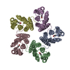
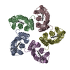
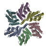
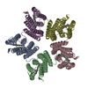
 ムービー
ムービー コントローラー


+ データを開く

- 基本情報
基本情報
| 登録情報 | データベース: PDB / ID: 7b03 | ||||||
|---|---|---|---|---|---|---|---|
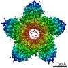
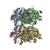
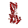
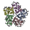
| タイトル | Cryo-EM structure of the green-light absorbing proteorhodopsin | ||||||
 要素 要素 | Proteorhodopsin | ||||||
 キーワード キーワード | PROTON TRANSPORT / Light-driven proton pump / microbial rhodopsin / retinal | ||||||
| 機能・相同性 |  機能・相同性情報 機能・相同性情報light-activated monoatomic ion channel activity / photoreceptor activity / phototransduction / plasma membrane 類似検索 - 分子機能 | ||||||
| 生物種 |  uncultured Gammaproteobacteria bacterium (環境試料) uncultured Gammaproteobacteria bacterium (環境試料) | ||||||
| 手法 | 電子顕微鏡法 / 単粒子再構成法 / クライオ電子顕微鏡法 / 解像度: 2.93 Å | ||||||
 データ登録者 データ登録者 | Hirschi, S. / Kalbermatter, D. / Fotiadis, D. | ||||||
| 資金援助 |  スイス, 1件 スイス, 1件
| ||||||
 引用 引用 | ジャーナル: Nat Commun / 年: 2021 タイトル: Cryo-EM structure and dynamics of the green-light absorbing proteorhodopsin. 著者: Stephan Hirschi / David Kalbermatter / Zöhre Ucurum / Thomas Lemmin / Dimitrios Fotiadis / 要旨: The green-light absorbing proteorhodopsin (GPR) is the archetype of bacterial light-driven proton pumps. Here, we present the 2.9 Å cryo-EM structure of pentameric GPR, resolving important ...The green-light absorbing proteorhodopsin (GPR) is the archetype of bacterial light-driven proton pumps. Here, we present the 2.9 Å cryo-EM structure of pentameric GPR, resolving important residues of the proton translocation pathway and the oligomerization interface. Superposition with the structure of a close GPR homolog and molecular dynamics simulations reveal conformational variations, which regulate the solvent access to the intra- and extracellular half channels harbouring the primary proton donor E109 and the proposed proton release group E143. We provide a mechanism for the structural rearrangements allowing hydration of the intracellular half channel, which are triggered by changing the protonation state of E109. Functional characterization of selected mutants demonstrates the importance of the molecular organization around E109 and E143 for GPR activity. Furthermore, we present evidence that helices involved in the stabilization of the protomer interfaces serve as scaffolds for facilitating the motion of the other helices. Combined with the more constrained dynamics of the pentamer compared to the monomer, these observations illustrate the previously demonstrated functional significance of GPR oligomerization. Overall, this work provides molecular insights into the structure, dynamics and function of the proteorhodopsin family that will benefit the large scientific community employing GPR as a model protein. | ||||||
| 履歴 |
|
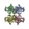
- 構造の表示
構造の表示
| ムービー |
ムービービューア |
|---|---|
| 構造ビューア | 分子: MolmilJmol/JSmol |
- ダウンロードとリンク
-ダウンロード
| PDBx/mmCIF形式 | 7b03.cif.gz | 190.8 KB | 表示 | PDBx/mmCIF形式 |
|---|---|---|---|---|
| PDB形式 | pdb7b03.ent.gz | 155.7 KB | 表示 | PDB形式 |
| PDBx/mmJSON形式 | 7b03.json.gz | ツリー表示 | PDBx/mmJSON形式 | |
| その他 |  その他のダウンロード その他のダウンロード |
-検証レポート
| 文書・要旨 | 7b03_validation.pdf.gz | 1 MB | 表示 | wwPDB検証レポート |
|---|---|---|---|---|
| 文書・詳細版 | 7b03_full_validation.pdf.gz | 1 MB | 表示 | |
| XML形式データ | 7b03_validation.xml.gz | 33.3 KB | 表示 | |
| CIF形式データ | 7b03_validation.cif.gz | 45.1 KB | 表示 | |
| アーカイブディレクトリ | https://data.pdbj.org/pub/pdb/validation_reports/b0/7b03ftp://data.pdbj.org/pub/pdb/validation_reports/b0/7b03 | HTTPS FTP |
-関連構造データ


-リンク
 PDBj
PDBj


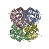
- 集合体
集合体
| 登録構造単位 | 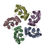
|
|---|---|
| 1 |
|
-要素
| #1: タンパク質 | 分子量: 25611.842 Da / 分子数: 5 / 由来タイプ: 組換発現 / 詳細: Expressed without signal sequence. 由来: (組換発現) uncultured Gammaproteobacteria bacterium (環境試料)発現宿主: #2: 化合物 | ChemComp-RET /   分子量: 284.436 Da / 分子数: 5 / 由来タイプ: 合成 / 式: C20H28O / タイプ: SUBJECT OF INVESTIGATION 分子量: 284.436 Da / 分子数: 5 / 由来タイプ: 合成 / 式: C20H28O / タイプ: SUBJECT OF INVESTIGATION研究の焦点であるリガンドがあるか | Y | Has protein modification | Y | |
|---|
-実験情報
-実験
| 実験 | 手法: 電子顕微鏡法 |
|---|---|
| EM実験 | 試料の集合状態: PARTICLE / 3次元再構成法: 単粒子再構成法 |
- 試料調製
試料調製
| 構成要素 | 名称: Pentamer of the green-light absorbing proteorhodopsin タイプ: COMPLEX / Entity ID: #1 / 由来: RECOMBINANT |
|---|---|
| 分子量 | 実験値: NO |
| 由来(天然) | 生物種: uncultured Gammaproteobacteria bacterium (環境試料) |
| 由来(組換発現) | 生物種: |
| 緩衝液 | pH: 7.5 |
| 試料 | 濃度: 3.5 mg/ml / 包埋: NO / シャドウイング: NO / 染色: NO / 凍結: YES |
| 試料支持 | グリッドの材料: COPPER / グリッドのタイプ: Quantifoil R1.2/1.3 |
| 急速凍結 | 装置: FEI VITROBOT MARK IV / 凍結剤: ETHANE / 湿度: 100 % / 凍結前の試料温度: 277 K |
- 電子顕微鏡撮影
| 実験機器 |  モデル: Titan Krios / 画像提供: FEI Company |
|---|---|
| 顕微鏡 | モデル: FEI TITAN KRIOS |
| 電子銃 | 電子線源:  FIELD EMISSION GUN / 加速電圧: 300 kV / 照射モード: SPOT SCAN FIELD EMISSION GUN / 加速電圧: 300 kV / 照射モード: SPOT SCAN |
| 電子レンズ | モード: BRIGHT FIELD |
| 撮影 | 電子線照射量: 1.36 e/Å2 / フィルム・検出器のモデル: GATAN K3 (6k x 4k) |
- 解析
解析
| EMソフトウェア |
| ||||||||||||||||||||||||
|---|---|---|---|---|---|---|---|---|---|---|---|---|---|---|---|---|---|---|---|---|---|---|---|---|---|
| CTF補正 | タイプ: PHASE FLIPPING AND AMPLITUDE CORRECTION | ||||||||||||||||||||||||
| 対称性 | 点対称性: C5 (5回回転対称) | ||||||||||||||||||||||||
| 3次元再構成 | 解像度: 2.93 Å / 解像度の算出法: FSC 0.143 CUT-OFF / 粒子像の数: 717107 詳細: Final map was obtained after applying density modification using Resolve Cryo-EM in Phenix. 対称性のタイプ: POINT |
