 Movie
Movie Controller


+ Open data

- Basic information
| Entry | Database: PDB / ID: 7axa | ||||||||||||
|---|---|---|---|---|---|---|---|---|---|---|---|---|---|
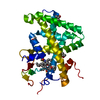
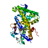
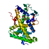
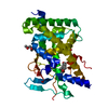
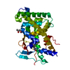
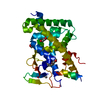
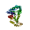
| Title | Crystal structure of the hPXR-LBD in complex with clotrimazole | ||||||||||||
 Components Components | Nuclear receptor subfamily 1 group I member 2 | ||||||||||||
 Keywords Keywords | NUCLEAR PROTEIN / NUCLEAR RECEPTOR HORMONE RECEPTOR PREGNANE X RECEPTOR | ||||||||||||
| Function / homology |  Function and homology information Function and homology informationcellular response to molecule of bacterial origin / intestinal epithelial structure maintenance / intermediate filament cytoskeleton / xenobiotic transport / steroid metabolic process / xenobiotic catabolic process / intracellular receptor signaling pathway / xenobiotic metabolic process / nuclear receptor binding / RNA polymerase II transcription regulatory region sequence-specific DNA binding ...cellular response to molecule of bacterial origin / intestinal epithelial structure maintenance / intermediate filament cytoskeleton / xenobiotic transport / steroid metabolic process / xenobiotic catabolic process / intracellular receptor signaling pathway / xenobiotic metabolic process / nuclear receptor binding / RNA polymerase II transcription regulatory region sequence-specific DNA binding / SUMOylation of intracellular receptors / Nuclear Receptor transcription pathway / nuclear receptor activity / sequence-specific double-stranded DNA binding / DNA-binding transcription activator activity, RNA polymerase II-specific / transcription regulator complex / DNA-binding transcription factor activity, RNA polymerase II-specific / cell differentiation / nuclear body / RNA polymerase II cis-regulatory region sequence-specific DNA binding / negative regulation of DNA-templated transcription / positive regulation of gene expression / regulation of DNA-templated transcription / chromatin / positive regulation of DNA-templated transcription / negative regulation of transcription by RNA polymerase II / signal transduction / positive regulation of transcription by RNA polymerase II / zinc ion binding / nucleoplasm / nucleus Similarity search - Function | ||||||||||||
| Biological species |  Homo sapiens (human) Homo sapiens (human) | ||||||||||||
| Method |  X-RAY DIFFRACTION / SYNCHROTRON / MOLECULAR REPLACEMENT / Resolution: 2.26 Å X-RAY DIFFRACTION / SYNCHROTRON / MOLECULAR REPLACEMENT / Resolution: 2.26 Å | ||||||||||||
 Authors Authors | Delfosse, V. / Granell, M. / Blanc, P. / Bourguet, W. | ||||||||||||
| Funding support |  France, 3items France, 3items
| ||||||||||||
 Citation Citation | Journal: Proc.Natl.Acad.Sci.USA / Year: 2021 Title: Mechanistic insights into the synergistic activation of the RXR-PXR heterodimer by endocrine disruptor mixtures. Authors: Delfosse, V. / Huet, T. / Harrus, D. / Granell, M. / Bourguet, M. / Gardia-Parege, C. / Chiavarina, B. / Grimaldi, M. / Le Mevel, S. / Blanc, P. / Huang, D. / Gruszczyk, J. / Demeneix, B. / ...Authors: Delfosse, V. / Huet, T. / Harrus, D. / Granell, M. / Bourguet, M. / Gardia-Parege, C. / Chiavarina, B. / Grimaldi, M. / Le Mevel, S. / Blanc, P. / Huang, D. / Gruszczyk, J. / Demeneix, B. / Cianferani, S. / Fini, J.B. / Balaguer, P. / Bourguet, W. | ||||||||||||
| History |
|
- Structure visualization
| Structure viewer | Molecule: MolmilJmol/JSmol |
|---|
- Downloads & links
-Download
| PDBx/mmCIF format | 7axa.cif.gz | 92.1 KB | Display | PDBx/mmCIF format |
|---|---|---|---|---|
| PDB format | pdb7axa.ent.gz | 55.3 KB | Display | PDB format |
| PDBx/mmJSON format | 7axa.json.gz | Tree view | PDBx/mmJSON format | |
| Others |  Other downloads Other downloads |
-Validation report
| Summary document | 7axa_validation.pdf.gz | 1.3 MB | Display | wwPDB validaton report |
|---|---|---|---|---|
| Full document | 7axa_full_validation.pdf.gz | 1.3 MB | Display | |
| Data in XML | 7axa_validation.xml.gz | 14.5 KB | Display | |
| Data in CIF | 7axa_validation.cif.gz | 20.5 KB | Display | |
| Arichive directory | https://data.pdbj.org/pub/pdb/validation_reports/ax/7axaftp://data.pdbj.org/pub/pdb/validation_reports/ax/7axa | HTTPS FTP |
-Related structure data
| Related structure data | 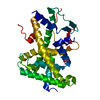 7ax8C  7ax9C  7axbC  7axcC  7axdC 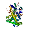 7axeC  7axfC  7axgC  7axhC  7axiC  7axjC  7axkC 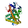 7axlC 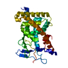 1ilgS S: Starting model for refinement C: citing same article ( |
|---|---|
| Similar structure data |


-Links
 PDBj
PDBj- Assembly
| Deposited unit | 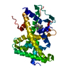
| ||||||||||||
|---|---|---|---|---|---|---|---|---|---|---|---|---|---|
| 1 |
| ||||||||||||
| 2 | 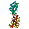
| ||||||||||||
| Unit cell |
|
-Components
| #1: Protein | Mass: 36747.465 Da / Num. of mol.: 1 Source method: isolated from a genetically manipulated source Source: (gene. exp.) Homo sapiens (human) / Gene: NR1I2, PXR / Plasmid: pET11 / Production host:  |
|---|---|
| #2: Chemical | ChemComp-CL6 /   Mass: 344.837 Da / Num. of mol.: 1 / Source method: obtained synthetically / Formula: C22H17ClN2 / Feature type: SUBJECT OF INVESTIGATION Mass: 344.837 Da / Num. of mol.: 1 / Source method: obtained synthetically / Formula: C22H17ClN2 / Feature type: SUBJECT OF INVESTIGATION |
| #3: Water | ChemComp-HOH /  Mass: 18.015 Da / Num. of mol.: 170 / Source method: isolated from a natural source / Formula: H2O Mass: 18.015 Da / Num. of mol.: 170 / Source method: isolated from a natural source / Formula: H2O |
| Has ligand of interest | Y |
-Experimental details
-Experiment
| Experiment | Method: X-RAY DIFFRACTION / Number of used crystals: 1 |
|---|
- Sample preparation
| Crystal | Density Matthews: 2.45 Å3/Da / Density % sol: 49.8 % |
|---|---|
| Crystal grow | Temperature: 291 K / Method: vapor diffusion, hanging drop / Details: 50 - 100 mM imidazole 8 - 14% isopropanol / PH range: 7.0 - 7.4 |
-Data collection
| Diffraction | Mean temperature: 100 K / Serial crystal experiment: N |
|---|---|
| Diffraction source | Source: SYNCHROTRON / Site: ESRF / Beamline: MASSIF-1 / Wavelength: 0.966 Å |
| Detector | Type: DECTRIS PILATUS3 2M / Detector: PIXEL / Date: Apr 18, 2018 |
| Radiation | Protocol: SINGLE WAVELENGTH / Monochromatic (M) / Laue (L): M / Scattering type: x-ray |
| Radiation wavelength | Wavelength: 0.966 Å / Relative weight: 1 |
| Reflection | Resolution: 2.26→45.68 Å / Num. obs: 17442 / % possible obs: 98.6 % / Redundancy: 7.7 % / Biso Wilson estimate: 35.57 Å2 / Rrim(I) all: 0.061 / Net I/σ(I): 23.95 |
| Reflection shell | Resolution: 2.26→2.32 Å / Mean I/σ(I) obs: 4.68 / Num. unique obs: 1184 / Rrim(I) all: 0.574 |
- Processing
| Software |
| |||||||||||||||||||||||||||||||||||||||||||||||||
|---|---|---|---|---|---|---|---|---|---|---|---|---|---|---|---|---|---|---|---|---|---|---|---|---|---|---|---|---|---|---|---|---|---|---|---|---|---|---|---|---|---|---|---|---|---|---|---|---|---|---|
| Refinement | Method to determine structure: MOLECULAR REPLACEMENT Starting model: 1ILG Resolution: 2.26→45.67 Å / SU ML: 0.2521 / Cross valid method: FREE R-VALUE / σ(F): 1.35 / Phase error: 24.8587 Stereochemistry target values: GeoStd + Monomer Library + CDL v1.2
| |||||||||||||||||||||||||||||||||||||||||||||||||
| Solvent computation | Shrinkage radii: 0.9 Å / VDW probe radii: 1.11 Å / Solvent model: FLAT BULK SOLVENT MODEL | |||||||||||||||||||||||||||||||||||||||||||||||||
| Displacement parameters | Biso mean: 41.31 Å2 | |||||||||||||||||||||||||||||||||||||||||||||||||
| Refinement step | Cycle: LAST / Resolution: 2.26→45.67 Å
| |||||||||||||||||||||||||||||||||||||||||||||||||
| Refine LS restraints |
| |||||||||||||||||||||||||||||||||||||||||||||||||
| LS refinement shell |
|
