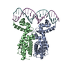
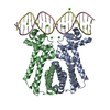
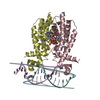
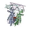
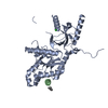
HTH-typetranscriptionalregulatorLuxR / LuxR family transcriptional regulator / Quorum sensing regulator LuxR / Quorum-sensing II master ...LuxR family transcriptional regulator / Quorum sensing regulator LuxR / Quorum-sensing II master regulator / TetR/AcrR family transcriptional regulator
Mass: 25304.752 Da / Num. of mol.: 2 Source method: isolated from a genetically manipulated source Source: (gene. exp.) Vibrio alginolyticus (bacteria) Gene: valR, luxR, AL539_16930, AOG25_16695, AT730_08150, K06K5_26160 Production host: Escherichia coli (E. coli) / References: UniProt: B4X9Q4
#2: DNA chain
DNA (5'-D(P*TP*AP*TP*AP*TP*AP*CP*AP*GP*TP*AP*AP*TP*GP*TP*CP*AP*TP*TP*AP*T)-3')
Mass: 6435.207 Da / Num. of mol.: 1 / Source method: obtained synthetically / Source: (synth.) Vibrio alginolyticus (bacteria)
#3: DNA chain
DNA (5'-D(P*AP*TP*AP*AP*TP*GP*AP*CP*AP*TP*TP*AP*CP*TP*GP*TP*AP*TP*AP*TP*A)-3')
Mass: 6444.222 Da / Num. of mol.: 1 / Source method: obtained synthetically / Source: (synth.) Vibrio alginolyticus (bacteria)
Has protein modification
Y
-
Experimental details
-
Experiment
Experiment
Method: X-RAY DIFFRACTION / Number of used crystals: 1
-
Sample preparation
Crystal
Density Matthews: 2.45 Å3/Da / Density % sol: 49.7 %
Crystal grow
Temperature: 291 K / Method: vapor diffusion, hanging drop Details: 50mM Sodium Formate, 50mM Tris PH7.5, 22% PEG 2000MME
-
Data collection
Diffraction
Mean temperature: 100 K / Serial crystal experiment: N
Diffraction source
Source: SYNCHROTRON / Site: ALBA / Beamline: XALOC / Wavelength: 0.9191 Å
In the structure databanks used in Yorodumi, some data are registered as the other names, "COVID-19 virus" and "2019-nCoV". Here are the details of the virus and the list of structure data.
Jan 31, 2019. EMDB accession codes are about to change! (news from PDBe EMDB page)
EMDB accession codes are about to change! (news from PDBe EMDB page)
The allocation of 4 digits for EMDB accession codes will soon come to an end. Whilst these codes will remain in use, new EMDB accession codes will include an additional digit and will expand incrementally as the available range of codes is exhausted. The current 4-digit format prefixed with “EMD-” (i.e. EMD-XXXX) will advance to a 5-digit format (i.e. EMD-XXXXX), and so on. It is currently estimated that the 4-digit codes will be depleted around Spring 2019, at which point the 5-digit format will come into force.
The EM Navigator/Yorodumi systems omit the EMD- prefix.
Related info.:Q: What is EMD? / ID/Accession-code notation in Yorodumi/EM Navigator
Yorodumi is a browser for structure data from EMDB, PDB, SASBDB, etc.
This page is also the successor to EM Navigator detail page, and also detail information page/front-end page for Omokage search.
The word "yorodu" (or yorozu) is an old Japanese word meaning "ten thousand". "mi" (miru) is to see.
Related info.:EMDB / PDB / SASBDB / Comparison of 3 databanks / Yorodumi Search / Aug 31, 2016. New EM Navigator & Yorodumi / Yorodumi Papers / Jmol/JSmol / Function and homology information / Changes in new EM Navigator and Yorodumi
 Movie
Movie Controller


 Open data
 Basic information
 Components
 Keywords
 Function and homology information
 Vibrio alginolyticus (bacteria)
 X-RAY DIFFRACTION /
  Authors
 Spain, 1items
  Citation
 Structure visualization
 Downloads & links
 Other downloads

 PDBj
PDBj


 Assembly
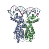
 Sample preparation
 Processing