 Movie
Movie Controller


+ Open data

- Basic information
| Entry | Database: PDB / ID: 7amb | |||||||||
|---|---|---|---|---|---|---|---|---|---|---|
| Title | Crystal structure of rsFolder2 in its fluorescent on-state | |||||||||
 Components Components | Green fluorescent protein | |||||||||
 Keywords Keywords | FLUORESCENT PROTEIN / GFP / Reversibly Photoswitchable Fluorescent Protein / Photoconvertible FP | |||||||||
| Function / homology | Green fluorescent protein, GFP / Green fluorescent protein-related / Green fluorescent protein / Green fluorescent protein / bioluminescence / generation of precursor metabolites and energy / Green fluorescent protein Function and homology information Function and homology information | |||||||||
| Biological species |   Aequorea victoria (jellyfish) Aequorea victoria (jellyfish) | |||||||||
| Method |  X-RAY DIFFRACTION / SYNCHROTRON / MOLECULAR REPLACEMENT / Resolution: 1.63 Å X-RAY DIFFRACTION / SYNCHROTRON / MOLECULAR REPLACEMENT / Resolution: 1.63 Å | |||||||||
 Authors Authors | Moreno-Chicano, T. / El Khatib, M. / Colletier, J.-P. | |||||||||
 Citation Citation | Journal: Chemphyschem / Year: 2022 Title: Rational Control of Off-State Heterogeneity in a Photoswitchable Fluorescent Protein Provides Switching Contrast Enhancement. Authors: Adam, V. / Hadjidemetriou, K. / Jensen, N. / Shoeman, R.L. / Woodhouse, J. / Aquila, A. / Banneville, A.S. / Barends, T.R.M. / Bezchastnov, V. / Boutet, S. / Byrdin, M. / Cammarata, M. / ...Authors: Adam, V. / Hadjidemetriou, K. / Jensen, N. / Shoeman, R.L. / Woodhouse, J. / Aquila, A. / Banneville, A.S. / Barends, T.R.M. / Bezchastnov, V. / Boutet, S. / Byrdin, M. / Cammarata, M. / Carbajo, S. / Eleni Christou, N. / Coquelle, N. / De la Mora, E. / El Khatib, M. / Moreno Chicano, T. / Bruce Doak, R. / Fieschi, F. / Foucar, L. / Glushonkov, O. / Gorel, A. / Grunbein, M.L. / Hilpert, M. / Hunter, M. / Kloos, M. / Koglin, J.E. / Lane, T.J. / Liang, M. / Mantovanelli, A. / Nass, K. / Nass Kovacs, G. / Owada, S. / Roome, C.M. / Schiro, G. / Seaberg, M. / Stricker, M. / Thepaut, M. / Tono, K. / Ueda, K. / Uriarte, L.M. / You, D. / Zala, N. / Domratcheva, T. / Jakobs, S. / Sliwa, M. / Schlichting, I. / Colletier, J.P. / Bourgeois, D. / Weik, M. | |||||||||
| History |
|
- Structure visualization
| Structure viewer | Molecule: MolmilJmol/JSmol |
|---|
- Downloads & links
-Download
| PDBx/mmCIF format | 7amb.cif.gz | 236.2 KB | Display | PDBx/mmCIF format |
|---|---|---|---|---|
| PDB format | pdb7amb.ent.gz | 191.4 KB | Display | PDB format |
| PDBx/mmJSON format | 7amb.json.gz | Tree view | PDBx/mmJSON format | |
| Others |  Other downloads Other downloads |
-Validation report
| Summary document | 7amb_validation.pdf.gz | 478.6 KB | Display | wwPDB validaton report |
|---|---|---|---|---|
| Full document | 7amb_full_validation.pdf.gz | 502.5 KB | Display | |
| Data in XML | 7amb_validation.xml.gz | 51.7 KB | Display | |
| Data in CIF | 7amb_validation.cif.gz | 74.8 KB | Display | |
| Arichive directory | https://data.pdbj.org/pub/pdb/validation_reports/am/7ambftp://data.pdbj.org/pub/pdb/validation_reports/am/7amb | HTTPS FTP |
-Related structure data
| Related structure data |  7amfC 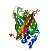 7o7cC  7o7dC  7o7eC 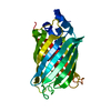 7o7hC  7o7uC 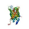 7o7vC  7o7wC 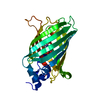 7o7xC 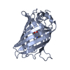 5dtzS S: Starting model for refinement C: citing same article ( |
|---|---|
| Similar structure data |
-Links
 PDBj
PDBj

- Assembly
| Deposited unit | 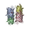
| ||||||||||||||||||
|---|---|---|---|---|---|---|---|---|---|---|---|---|---|---|---|---|---|---|---|
| 1 | 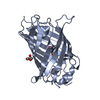
| ||||||||||||||||||
| 2 | 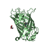
| ||||||||||||||||||
| 3 | 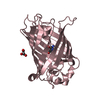
| ||||||||||||||||||
| 4 | 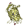
| ||||||||||||||||||
| Unit cell |
| ||||||||||||||||||
| Components on special symmetry positions |
|

-Components
| #1: Protein | Mass: 26724.070 Da / Num. of mol.: 4 Mutation: S30R, Y39N, F64L, S65A, Q69L, Q80R, F99S, N105T, M153T, V163S, I171V, A206K Source method: isolated from a genetically manipulated source Source: (gene. exp.) Aequorea victoria (jellyfish) / Gene: GFPProduction host:  References: UniProt: P42212 #2: Chemical | ChemComp-GOL /   Mass: 92.094 Da / Num. of mol.: 6 / Source method: obtained synthetically / Formula: C3H8O3 Mass: 92.094 Da / Num. of mol.: 6 / Source method: obtained synthetically / Formula: C3H8O3#3: Water | ChemComp-HOH / |  Mass: 18.015 Da / Num. of mol.: 1033 / Source method: isolated from a natural source / Formula: H2O Mass: 18.015 Da / Num. of mol.: 1033 / Source method: isolated from a natural source / Formula: H2OHas ligand of interest | Y | |
|---|
-Experimental details
-Experiment
| Experiment | Method: X-RAY DIFFRACTION / Number of used crystals: 1 |
|---|
- Sample preparation
| Crystal | Density Matthews: 2.21 Å3/Da / Density % sol: 44 % / Description: Large thick plates |
|---|---|
| Crystal grow | Temperature: 293 K / Method: vapor diffusion, hanging drop / pH: 8.5 / Details: 20% PEG 3350, 100 mM Tris pH 8.5, 20 mM NaCl |
-Data collection
| Diffraction | Mean temperature: 100 K / Serial crystal experiment: N |
|---|---|
| Diffraction source | Source: SYNCHROTRON / Site: ESRF  / Beamline: MASSIF-3 / Wavelength: 0.97 Å / Beamline: MASSIF-3 / Wavelength: 0.97 Å |
| Detector | Type: DECTRIS PILATUS3 2M / Detector: PIXEL / Date: Jul 1, 2015 |
| Radiation | Protocol: SINGLE WAVELENGTH / Monochromatic (M) / Laue (L): M / Scattering type: x-ray |
| Radiation wavelength | Wavelength: 0.97 Å / Relative weight: 1 |
| Reflection | Resolution: 1.63→47.98 Å / Num. obs: 115478 / % possible obs: 99.7 % / Redundancy: 3.5 % / Biso Wilson estimate: 18.43 Å2 / CC1/2: 0.999 / Rrim(I) all: 0.065 / Χ2: 0.98 / Net I/σ(I): 11 |
| Reflection shell | Resolution: 1.63→1.66 Å / Redundancy: 3.2 % / Mean I/σ(I) obs: 1.6 / Num. unique obs: 5788 / CC1/2: 0.747 / Rrim(I) all: 0.73 / Χ2: 0.99 / % possible all: 99.7 |
- Processing
| Software |
| ||||||||||||||||||||||||||||||||||||||||||||||||||||||||||||
|---|---|---|---|---|---|---|---|---|---|---|---|---|---|---|---|---|---|---|---|---|---|---|---|---|---|---|---|---|---|---|---|---|---|---|---|---|---|---|---|---|---|---|---|---|---|---|---|---|---|---|---|---|---|---|---|---|---|---|---|---|---|
| Refinement | Method to determine structure: MOLECULAR REPLACEMENT Starting model: 5dtz Resolution: 1.63→47.98 Å / Cor.coef. Fo:Fc: 0.978 / Cor.coef. Fo:Fc free: 0.966 / SU B: 2.295 / SU ML: 0.074 / Cross valid method: THROUGHOUT / σ(F): 0 / ESU R: 0.086 / ESU R Free: 0.089 / Stereochemistry target values: MAXIMUM LIKELIHOOD Details: HYDROGENS HAVE BEEN ADDED IN THE RIDING POSITIONS U VALUES : REFINED INDIVIDUALLY
| ||||||||||||||||||||||||||||||||||||||||||||||||||||||||||||
| Solvent computation | Ion probe radii: 0.8 Å / Shrinkage radii: 0.8 Å / VDW probe radii: 1.2 Å / Solvent model: MASK | ||||||||||||||||||||||||||||||||||||||||||||||||||||||||||||
| Displacement parameters | Biso max: 92.55 Å2 / Biso mean: 24.127 Å2 / Biso min: 11.24 Å2
| ||||||||||||||||||||||||||||||||||||||||||||||||||||||||||||
| Refinement step | Cycle: final / Resolution: 1.63→47.98 Å
| ||||||||||||||||||||||||||||||||||||||||||||||||||||||||||||
| Refine LS restraints |
| ||||||||||||||||||||||||||||||||||||||||||||||||||||||||||||
| LS refinement shell | Resolution: 1.63→1.672 Å / Rfactor Rfree error: 0 / Total num. of bins used: 20
|
