 Movie
Movie Controller


+ Open data

- Basic information
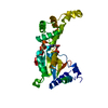
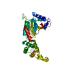
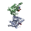
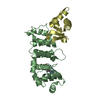
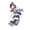
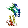
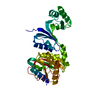
| Entry | Database: PDB / ID: 7aed | |||||||||
|---|---|---|---|---|---|---|---|---|---|---|
| Title | VirB8 domain of PrgL from Enterococcus faecalis pCF10 | |||||||||
 Components Components | PrgL | |||||||||
 Keywords Keywords | MEMBRANE PROTEIN / conjugation / T4SS / VirB8 | |||||||||
| Function / homology | membrane / PrgL Function and homology information Function and homology information | |||||||||
| Biological species |   Enterococcus faecalis (bacteria) Enterococcus faecalis (bacteria) | |||||||||
| Method |  X-RAY DIFFRACTION / SYNCHROTRON / SIRAS / SAD / Resolution: 1.753 Å X-RAY DIFFRACTION / SYNCHROTRON / SIRAS / SAD / Resolution: 1.753 Å | |||||||||
 Authors Authors | Jaeger, F. / Berntsson, R.P.A. | |||||||||
| Funding support |  Sweden, 2items Sweden, 2items
| |||||||||
 Citation Citation | Journal: Structure / Year: 2022 Title: Structure of the enterococcal T4SS protein PrgL reveals unique dimerization interface in the VirB8 protein family. Authors: Jager, F. / Lamy, A. / Sun, W.S. / Guerini, N. / Berntsson, R.P. #1: Journal: Biorxiv / Year: 2020Title: Structure of the enterococcal T4SS protein PrgL reveals unique dimerization interface in the VirB8 protein family Authors: Jaeger, F. / Lamy, A. / Guerini, N. / Sun, W.S. / Berntsson, R.P.A. | |||||||||
| History |
|
- Structure visualization
| Structure viewer | Molecule: MolmilJmol/JSmol |
|---|
- Downloads & links
-Download
| PDBx/mmCIF format | 7aed.cif.gz | 73 KB | Display | PDBx/mmCIF format |
|---|---|---|---|---|
| PDB format | pdb7aed.ent.gz | 52.9 KB | Display | PDB format |
| PDBx/mmJSON format | 7aed.json.gz | Tree view | PDBx/mmJSON format | |
| Others |  Other downloads Other downloads |
-Validation report
| Arichive directory | https://data.pdbj.org/pub/pdb/validation_reports/ae/7aedftp://data.pdbj.org/pub/pdb/validation_reports/ae/7aed | HTTPS FTP |
|---|
-Related structure data
| Similar structure data |
|---|


-Links
 PDBj
PDBj- Assembly
| Deposited unit | 
| ||||||||
|---|---|---|---|---|---|---|---|---|---|
| 1 |
| ||||||||
| Unit cell |
|
-Components
| #1: Protein | Mass: 22541.057 Da / Num. of mol.: 2 Source method: isolated from a genetically manipulated source Source: (gene. exp.) Enterococcus faecalis (bacteria) / Gene: prgL / Production host: Lactococcus lactis (lactic acid bacteria) / References: UniProt: D1LHF8#2: Chemical |   Mass: 209.240 Da / Num. of mol.: 2 / Source method: obtained synthetically / Formula: C8H19NO5 / Comment: pH buffer*YM Mass: 209.240 Da / Num. of mol.: 2 / Source method: obtained synthetically / Formula: C8H19NO5 / Comment: pH buffer*YM#3: Water | ChemComp-HOH / |  Mass: 18.015 Da / Num. of mol.: 155 / Source method: isolated from a natural source / Formula: H2O Mass: 18.015 Da / Num. of mol.: 155 / Source method: isolated from a natural source / Formula: H2OHas ligand of interest | N | |
|---|
-Experimental details
-Experiment
| Experiment | Method: X-RAY DIFFRACTION / Number of used crystals: 1 |
|---|
- Sample preparation
| Crystal | Density Matthews: 2.2 Å3/Da / Density % sol: 44.05 % |
|---|---|
| Crystal grow | Temperature: 293 K / Method: vapor diffusion, sitting drop / pH: 5.5 / Details: bis-Tris, PEG 3350 |
-Data collection
| Diffraction | Mean temperature: 100 K / Serial crystal experiment: N |
|---|---|
| Diffraction source | Source: SYNCHROTRON / Site: MAX IV / Beamline: BioMAX / Wavelength: 0.9793 Å |
| Detector | Type: DECTRIS EIGER X 16M / Detector: PIXEL / Date: Jun 28, 2019 |
| Radiation | Protocol: SINGLE WAVELENGTH / Monochromatic (M) / Laue (L): M / Scattering type: x-ray |
| Radiation wavelength | Wavelength: 0.9793 Å / Relative weight: 1 |
| Reflection | Resolution: 1.753→41.86 Å / Num. obs: 76794 / % possible obs: 99.6 % / Redundancy: 5.3 % / CC1/2: 0.99 / Rrim(I) all: 0.05 / Net I/σ(I): 16.3 |
| Reflection shell | Resolution: 1.753→1.86 Å / Num. unique obs: 12242 / CC1/2: 0.51 |
-Phasing
| Phasing | Method: SAD |
|---|
- Processing
| Software |
| ||||||||||||||||||||||||||||||||||||||||||||||||||||||||||||||||||||||||||||||||||||||||||||||||
|---|---|---|---|---|---|---|---|---|---|---|---|---|---|---|---|---|---|---|---|---|---|---|---|---|---|---|---|---|---|---|---|---|---|---|---|---|---|---|---|---|---|---|---|---|---|---|---|---|---|---|---|---|---|---|---|---|---|---|---|---|---|---|---|---|---|---|---|---|---|---|---|---|---|---|---|---|---|---|---|---|---|---|---|---|---|---|---|---|---|---|---|---|---|---|---|---|---|
| Refinement | Method to determine structure: SIRAS / Resolution: 1.753→34.74 Å / SU ML: 0.2 / Cross valid method: THROUGHOUT / σ(F): 1.96 / Phase error: 21.13 / Stereochemistry target values: ML
| ||||||||||||||||||||||||||||||||||||||||||||||||||||||||||||||||||||||||||||||||||||||||||||||||
| Solvent computation | Shrinkage radii: 0.9 Å / VDW probe radii: 1.11 Å / Solvent model: FLAT BULK SOLVENT MODEL | ||||||||||||||||||||||||||||||||||||||||||||||||||||||||||||||||||||||||||||||||||||||||||||||||
| Displacement parameters | Biso max: 106.44 Å2 / Biso mean: 45.4746 Å2 / Biso min: 24.75 Å2 | ||||||||||||||||||||||||||||||||||||||||||||||||||||||||||||||||||||||||||||||||||||||||||||||||
| Refinement step | Cycle: final / Resolution: 1.753→34.74 Å
| ||||||||||||||||||||||||||||||||||||||||||||||||||||||||||||||||||||||||||||||||||||||||||||||||
| LS refinement shell | Refine-ID: X-RAY DIFFRACTION / Rfactor Rfree error: 0
|
