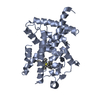
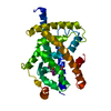
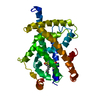
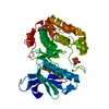
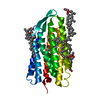
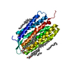
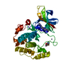
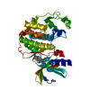
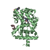
Entry Database : PDB / ID : 7a7hTitle Crystal structure of PPARgamma in complex with compound TK90 Peroxisome proliferator-activated receptor gamma Keywords / / / / Function / homology Function Domain/homology Component
/ / / / / / / / / / / / / / / / / / / / / / / / / / / / / / / / / / / / / / / / / / / / / / / / / / / / / / / / / / / / / / / / / / / / / / / / / / / / / / / / / / / / / / / / / / / / / / / / / / / / / / / / / / / / / / / / / / / / / / / / / / / / / / / / / / / / / Biological species Homo sapiens (human)Method / / / Resolution : 2.4 Å Authors Ni, X. / Kirchner, T. / Proschak, E. / Chaikuad, A. / Knapp, S. / Structural Genomics Consortium (SGC) Journal : J.Med.Chem. / Year : 2021Title : Combined Cardioprotective and Adipocyte Browning Effects Promoted by the Eutomer of Dual sEH/PPAR gamma Modulator.Authors: Hartmann, M. / Bibli, S.I. / Tews, D. / Ni, X. / Kircher, T. / Kramer, J.S. / Kilu, W. / Heering, J. / Hernandez-Olmos, V. / Weizel, L. / Scriba, G.K.E. / Krait, S. / Knapp, S. / Chaikuad, A. ... Authors : Hartmann, M. / Bibli, S.I. / Tews, D. / Ni, X. / Kircher, T. / Kramer, J.S. / Kilu, W. / Heering, J. / Hernandez-Olmos, V. / Weizel, L. / Scriba, G.K.E. / Krait, S. / Knapp, S. / Chaikuad, A. / Merk, D. / Fleming, I. / Fischer-Posovszky, P. / Proschak, E. History Deposition Aug 28, 2020 Deposition site / Processing site Revision 1.0 Aug 4, 2021 Provider / Type Revision 1.1 Jan 31, 2024 Group / Database references / Refinement descriptionCategory chem_comp_atom / chem_comp_bond ... chem_comp_atom / chem_comp_bond / database_2 / pdbx_initial_refinement_model Item / _database_2.pdbx_database_accession
Show all Show less
 Movie
Movie Controller


 Open data
 Basic information
 Components
 Keywords
 Function and homology information
 Homo sapiens (human)
 X-RAY DIFFRACTION /
  Authors
 Citation
 Structure visualization
 Downloads & links
 Other downloads
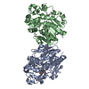


 PDBj
PDBj Assembly


 Mass: 409.399 Da / Num. of mol.: 1 / Source method: obtained synthetically / Formula: C21H22F3NO4 / Feature type: SUBJECT OF INVESTIGATION
 Mass: 62.068 Da / Num. of mol.: 1 / Source method: obtained synthetically / Formula: C2H6O2
 Mass: 18.015 Da / Num. of mol.: 20 / Source method: isolated from a natural source / Formula: H2O
 Sample preparation
 / Beamline: X06DA / Wavelength: 1 Å
 Processing